

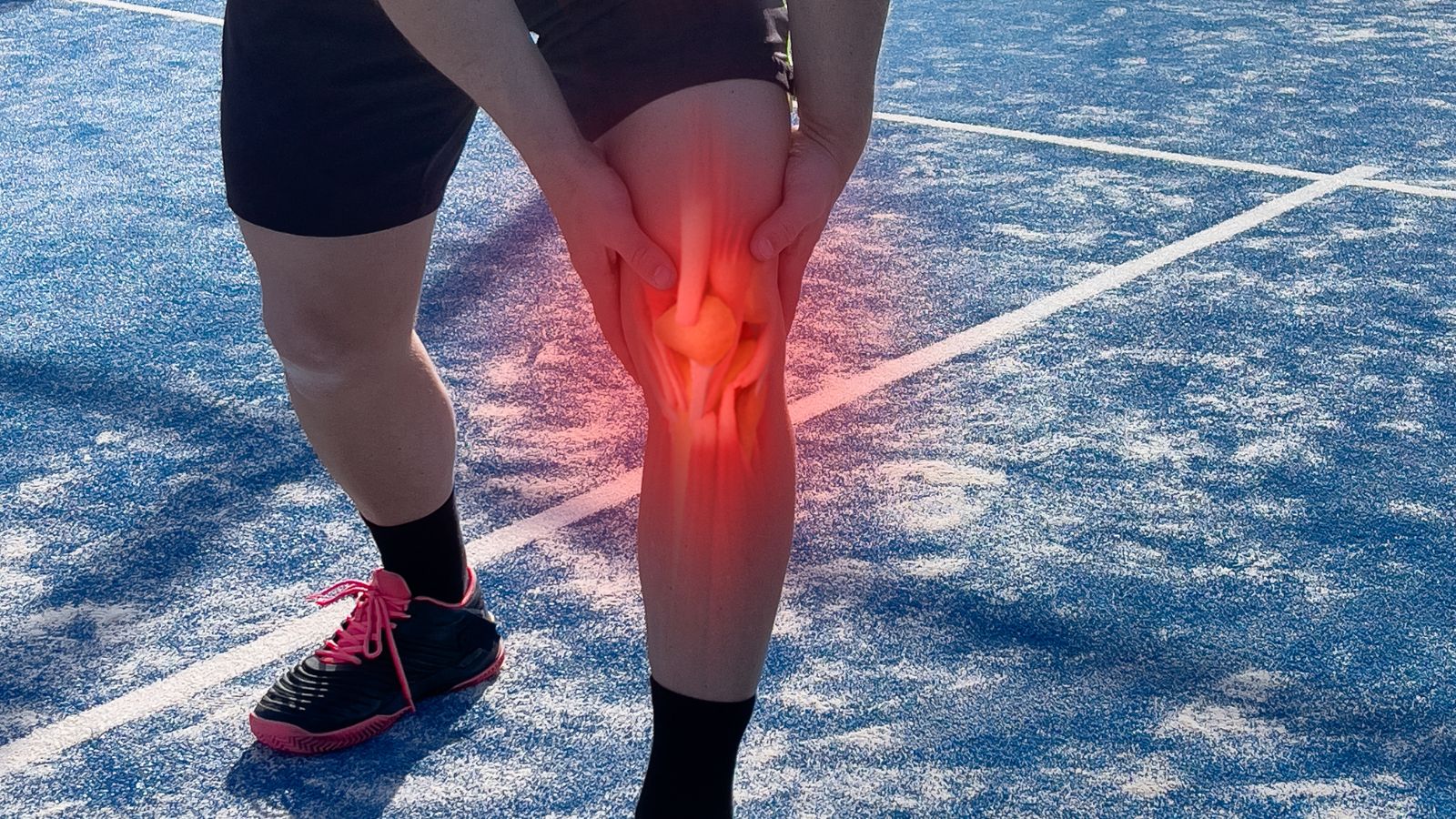
Prof. Dr. Murat Demirel, one of the best orthopedic doctors in Ankara specializing in the treatment of chondromalacia patella (runner’s knee), stands out with his expertise and experience in managing this condition caused by the softening or damage of the cartilage tissue under the kneecap. Chondromalacia patella is especially common among athletes, individuals engaged in intense physical activity, and those working in professions that put strain on the knee joint. The condition manifests itself with knee pain, difficulty climbing stairs, and discomfort when sitting for long periods. Prof. Dr. Demirel provides personalized treatment plans for each patient at hospitals in Ankara with advanced medical equipment and high hygiene standards.
Treatment options include rest, medication, physical therapy practices, and modern methods such as PRP or hyaluronic acid injections. In necessary cases, arthroscopic surgical interventions may also be performed. After chondromalacia patella treatment, following medical advice ensures a fast, safe, and lasting recovery. In addition, transparent information about chondromalacia patella treatment costs in Ankara is provided to help patients make informed decisions. You can also protect your knee health, reduce your pain, and return to your active life by contacting us to book an appointment.
| Disease Name | Chondromalacia Patella (Runner’s Knee) |
| Affected Area | Patellofemoral joint (cartilage surface beneath the kneecap) |
| Symptoms | Anterior knee pain, pain when climbing or descending stairs, discomfort when sitting for long periods, clicking or grinding sensation in the knee |
| Diagnostic Methods | Physical examination, MRI imaging, arthroscopy |
| Causes | Cartilage softening and degeneration; overuse, muscle imbalances, misalignment of the knee |
| Risk Factors | Young age, female gender, runners and athletes, flat feet, muscle imbalances |
| Treatment Methods | Conservative: rest, ice, NSAIDs, quadriceps strengthening exercises, knee brace, physical therapy |
| Surgical Options | Arthroscopic debridement, lateral release, tibial tubercle translocation (in advanced cases) |
| Complications | Chronic pain, reduced knee function, progressive cartilage damage |
| Recovery Process | A few weeks to several months with conservative treatment; 3–6 months after surgery |
| Prevention Methods | Wearing appropriate shoes, warming up before exercise, strengthening muscles around the knee, choosing proper running surfaces |
| Follow-Up | Monitoring physical therapy progress, long-term exercise programs to prevent recurrences |

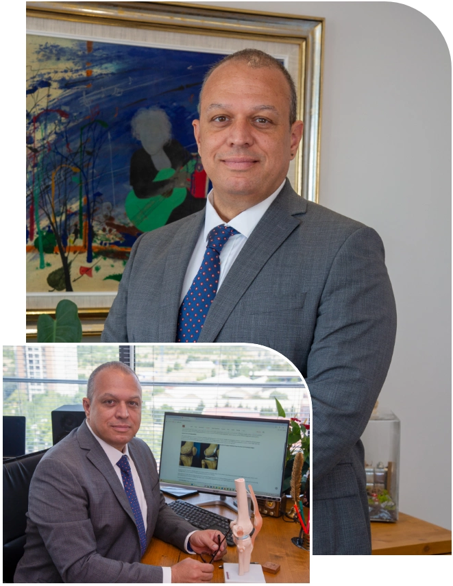
Prof. Dr. Murat Demirel
Orthopedics and Traumatology Specialist
Orthopedics Specialist Prof. Dr. Murat Demirel was born in Ankara in 1974. He completed his primary education at Ankara Kavaklıdere Primary School and his secondary and high school education at Ankara Atatürk Anatolian High School. Dr. Demirel graduated from Ankara University Faculty of Medicine in 1998 and completed his residency in Orthopedics and Traumatology at Ankara Numune Training and Research Hospital, 1st Orthopedics and Traumatology Clinic, in 2004.
PhD
Ankara University Institute of Health Sciences
Specialization
Ankara Numune Training and Research Hospital, 1st Orthopedics Clinic
Medical School
Ankara University Faculty of Medicine
Yazı İçeriği
What is Chondromalacia Patella?
When stripped of medical jargon, Chondromalacia Patella essentially means the softening and deterioration of the articular cartilage covering the back surface of the kneecap (patella). To better understand this, we can use a simple analogy: imagine the cartilage under your kneecap as the smooth, slick surface of a brand-new non-stick pan. Thanks to this surface, your kneecap glides effortlessly in a special groove on the thigh bone (trochlear groove) as you move.
When Chondromalacia Patella begins, small scratches, roughness, and softening develop on this once-smooth surface. It is no longer perfectly even. This increases friction, causes catching with each kneecap movement, and eventually leads to the well-known discomforting pain. In other words, the problem stems from a disruption in the perfect mechanics of the knee joint. It should not be considered merely a “cartilage disease,” because this condition is influenced by many factors, from muscle balance controlling the kneecap, to foot structure, and even the anatomy of the pelvis.
What are the symptoms of Chondromalacia Patella?
Chondromalacia Patella usually develops gradually, and its symptoms can vary from person to person. However, there are some typical signals commonly described by patients. Recognizing these signs is the first step toward early prevention. The most common symptoms are:
- A dull, aching pain felt in the front, back, or sides of the kneecap
- Increased pain when climbing or descending stairs (especially descending)
- Difficulty with squatting or activities requiring knee bending
- Pain and stiffness when standing up after prolonged sitting (Theater Sign)
- Grinding, creaking, or popping sounds from the knee (Crepitus)
- Occasional mild swelling, especially after strenuous activity
- A general sense of instability or giving way in the knee
- Gradual weakness in the front thigh muscle (quadriceps)
The most classic sign is what we call the “theater sign.” During a long movie or trip, when your knee remains bent, pressure inside the kneecap joint increases. The already sensitive cartilage cannot withstand this pressure, and the first step you take upon standing is accompanied by sharp pain. Similarly, the “crunching” sounds from the knee, known as crepitus, often worry patients. These sounds occur as the roughened cartilage surfaces rub against each other. On their own, without pain, they are not considered dangerous. However, if accompanied by pain, it is an important indicator that it’s time to consult a specialist.
Why does Chondromalacia Patella occur and who is at higher risk?
It is almost impossible to attribute Chondromalacia Patella to a single cause. Like puzzle pieces, several factors often combine to create this condition. Understanding these factors guides both prevention and treatment strategies. Let’s look at them more closely.
Kneecap Misalignment: Perhaps the most important cause. The kneecap should move centrally within the groove on the thigh bone, like a train on rails. But due to muscle imbalances or anatomical reasons, if the kneecap shifts to one side, excessive pressure is placed on a specific part of the cartilage. Over time, this uneven load wears down that area.
Muscle Imbalances: Imagine a “tug of war” between the muscles surrounding the kneecap. The inner thigh muscle (VMO) and the outer thigh structures (Vastus Lateralis and IT Band) pull the kneecap in different directions. If the outer structures are too tight and strong, and the inner VMO is weak, the outer side wins and constantly pulls the kneecap outward. This causes misalignment and friction. Similarly, weak hip and core muscles can disrupt overall leg mechanics, causing the knee to collapse inward and increasing stress on the kneecap.
Overuse and Training Errors: Sports like running, cycling, or jumping put repetitive stress on the knee and can cause micro-traumas in the cartilage. Increasing training intensity or duration without preparation, running downhill, or exercising on hard surfaces are significant risk factors.
Anatomical and Biomechanical Factors: Some people are naturally predisposed to this condition. Risk factors include:
- Being female (due to a wider pelvis and different musculoskeletal geometry)
- Flat feet (pes planus)
- Foot problems like inward or outward rolling
- Leg deformities such as knock knees (valgus) or bowlegs (varus)
- A shallow groove in the thigh bone (trochlear dysplasia)
- Previous knee injuries (direct trauma, fractures, or dislocations)
- Excess weight (increasing the load on the knees)
Contact us for detailed information and an appointment!
How is cartilage damage staged in Chondromalacia Patella?
To understand the severity of cartilage damage and shape the treatment plan accordingly, a staging system is used. This acts like a roadmap, showing us how deep the damage has progressed. One of the most widely used systems is the Outerbridge Classification, which typically describes the damage in four stages.
These stages are:
Stage 1: Cartilage softening and swelling only. The surface is still smooth but has lost its normal firmness. This stage is usually reversible.
Stage 2: Small cracks and roughness appear on the cartilage surface. The damage has penetrated less than half of the cartilage thickness.
Stage 3: The cracks are now much deeper, reaching the underlying bone tissue. The cartilage surface may appear “like crab meat.” The damage affects more than half of the cartilage thickness.
Stage 4: This is the most advanced stage. The cartilage in that area is completely worn away, exposing the bone underneath. This can cause the bones to rub against each other.
This staging is usually performed with MRI (Magnetic Resonance Imaging) or direct observation during arthroscopic surgery. The most important point to know is that damage detected in early stages (especially Stage 1 and 2) can be stopped and symptoms largely eliminated with proper treatment and lifestyle changes.
What steps are taken to diagnose Chondromalacia Patella?
The path to correct treatment always begins with an accurate diagnosis. In diagnosing Chondromalacia Patella, we do not rely on a single finding or test; instead, we piece together clues like a detective, making a holistic assessment. This process usually consists of three main steps: listening to you, examining you, and using imaging methods.
Everything begins with your story. From the moment you walk in, every detail you share is a clue for us.
When did your pain start?
Is there a specific movement or activity that triggers the pain?
What is the character of the pain (burning, stabbing, aching)?
Do you feel catching, locking, or giving way in your knee?
Do you have typical signs such as the “theater sign”?
Have you ever had a knee injury before?
What sports do you play or what is your profession?
The answers to these questions provide valuable preliminary information and shape our examination.
Physical Examination: After listening to you, we move on to assessing the problem with our eyes and hands. We observe everything from your standing posture to your gait. We look at the overall alignment of your legs and how your feet strike the ground. On the exam table, we test your knee’s range of motion, the strength and flexibility of your muscles (especially the quadriceps and hip muscles). We evaluate kneecap movement with special maneuvers and check for sensitive points and friction sounds (crepitus). This examination plays a critical role in uncovering underlying biomechanical problems such as muscle imbalances or misalignments.
Imaging Methods: To confirm our clinical findings and rule out other possible issues (such as meniscus tears or ligament injuries), we use imaging methods.
X-ray: Usually the first test requested. It does not directly show cartilage but allows us to evaluate bone structure, kneecap position, and possible arthritis findings.
Magnetic Resonance Imaging (MRI): This is the gold standard for diagnosing Chondromalacia Patella. MRI allows us to see cartilage, soft tissues, and bone marrow in detail. It clearly shows cartilage softening, swelling, cracks, and the stage of the damage.
What are the non-surgical treatment options for Chondromalacia Patella?
As we always tell our patients, the strongest weapon in the battle against Chondromalacia Patella is non-surgical, conservative treatment methods. The vast majority of cases can improve without surgery with patience and the right approach. This treatment is not a single magic wand but a combination of different strategies working together. The following methods form the foundation of your healing journey:
Activity Modification: The first rule is to temporarily avoid movements that trigger pain. Stopping activities that put strain on the kneecap, such as kneeling, deep squatting, jumping, or running downhill, gives the cartilage a chance to rest and heal. This does not mean quitting sports entirely; it means adjusting them wisely. For example, switching from running to swimming or elliptical cycling can be a good alternative.
Ice and Rest: During periods of acute pain and swelling, applying ice to the affected area for 15–20 minutes several times a day is very effective in reducing inflammation and pain. Resting with the knee elevated above heart level also helps reduce swelling.
Medication: Anti-inflammatory drugs or creams prescribed by your doctor can be used to control pain and inflammation. These medications are not the cure themselves; they simply provide comfort during the healing process and allow you to participate in physical therapy more comfortably.
Physical Therapy and Exercise: This is the heart of conservative treatment. As we will detail in the next section, proper exercises address the root of the problem and offer a permanent solution.
Supportive Equipment:
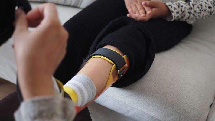
Knee Braces or Taping: Special taping techniques (such as McConnell taping) or braces can help the kneecap move along the correct track and reduce pain during activity. These are like “crutches,” supporting you during recovery.
Custom Insoles (Orthotics): If the underlying problem is flat feet, proper insole use can correct overall leg mechanics and eliminate abnormal load on the kneecap.
Weight Control: Every extra kilogram of body weight puts 3–4 times more load on the knees while walking, and even more when climbing stairs. Reaching and maintaining a healthy weight is one of the most important investments you can make to protect your knees.
What is the role of physical therapy and exercises in Chondromalacia Patella treatment?
If we compare Chondromalacia Patella treatment to a building, physical therapy and exercises are definitely the foundation. All other methods are built upon this foundation. Our goal is not only to relieve pain but to resolve the biomechanical problem causing the pain at its root. This is where properly planned chondromalacia patella exercises come into play, opening the door to lasting recovery.
The main goals of physical therapy are:
Balanced Muscle Strengthening: The problem is often not just in the knee. Strengthening weak hip and core muscles, which cause incorrect kneecap movement, is as important, if not more, than strengthening the muscles around the knee. Strong hip muscles bear body weight better and reduce stress on the knee. Targeted strengthening of the inner quadriceps muscle (VMO) helps keep the kneecap on track.
Stretching Tight Structures: Tight outer thigh structures (iliotibial band) and hamstring muscles can worsen the problem by pulling the kneecap outward. Regularly stretching these groups allows freer and more correct kneecap movement.
Relearning Correct Movement Patterns: With the help of a physiotherapist, you relearn how to perform daily movements like squatting and walking with proper biomechanics. This is vital to preventing recurrence of the problem.
Recommended Exercise Types:
- Isometric quadriceps contractions (tightening without changing muscle length)
- Straight leg raises
- Hip-targeting exercises (bridges, side leg raises, clamshells)
- Low-resistance stationary bike with high seat setting
- Water exercises (water buoyancy reduces knee load)
- Mini squats within a pain-free range (wall slides)
- Activities to Avoid or Perform with Caution:
- Deep squats and lunges
- Leg extension machine
- High-impact sports involving jumping (volleyball, basketball)
- Running downhill or on hard surfaces
- Stair climbing machines (Stairmaster)
Remember, every patient is different, and an exercise beneficial for one person may harm another. Therefore, instead of following advice from the internet or friends, you must follow a program specifically designed for you by a specialist to ensure treatment success.
When is surgery needed for Chondromalacia Patella?
One of our patients’ biggest concerns is whether this condition will inevitably require surgery. I can reassure you on this: in treating Chondromalacia Patella, surgery is always our last option. Surgical decision-making only comes into play when all other methods have been tried and failed, under specific and clear conditions.
The situations that may lead us to consider surgery are:
- No significant improvement in pain despite at least 6 months of consistent non-surgical treatment (physical therapy, medications, activity modification, etc.).
- Pain so severe and persistent that it prevents the patient from performing daily activities (such as climbing stairs, sitting and standing), seriously lowering quality of life.
- Imaging studies such as MRI show a detached piece of cartilage floating freely within the joint (loose body).
- Cartilage damage accompanied by a significant anatomical problem that requires surgical correction (for example, a ligament tear causing recurrent kneecap dislocations or a severe alignment problem).
- Advanced stage (Stage 4), with deep cartilage damage reaching the bone and covering a large area, especially in young and active patients, requiring cartilage repair techniques.
What is the recovery process after Chondromalacia Patella surgery?
The success of surgery for Chondromalacia Patella depends not only on the procedure performed in the operating room but equally on the postoperative rehabilitation process. This is essentially a second phase of treatment and requires the patient’s patience, compliance, and determination. The recovery process varies greatly depending on the type of surgery performed.
After Arthroscopic Debridement (Shaving): This is usually the simplest procedure with the fastest recovery. In the first few weeks after surgery, it is important to control swelling in the knee and avoid stressing the joint. Patients typically use crutches for a short time and quickly begin physical therapy. Low-impact exercises are introduced first, followed gradually by a return to normal activities and sports.
After Microfracture or Cartilage Repair Procedures (ACI, OATS): These cartilage-regeneration surgeries are more complex and require a much longer and more careful rehabilitation period.
Non-Weight-Bearing Period: Patients usually need to avoid putting weight on the operated leg, or put very limited weight, for 6 to 8 weeks to allow the repaired cartilage tissue to heal and integrate. Crutches are mandatory during this time.
Continuous Passive Motion (CPM) Machine: Sometimes devices called CPM are used, which move the knee in a controlled manner without the patient using active muscle power, in order to maintain joint mobility and promote cartilage nutrition.
Prolonged and Gradual Physical Therapy: Once weight-bearing begins, a very cautious and slow physical therapy program starts. Regaining muscle strength, balance, and endurance can take months.
Return to Sports: After these major surgeries, returning to high-impact sports such as running and jumping usually takes 9 months to 1 year, sometimes even longer.
Frequently Asked Questions
What is Chondromalacia Patella (Runner’s Knee)?
Chondromalacia Patella is a knee condition caused by the softening and damage of the cartilage tissue behind the kneecap. It is most common in young and active individuals, especially runners and athletes.
What causes Runner’s Knee?
The most common causes of Chondromalacia Patella are excessive stress on the kneecap, incorrect exercise techniques, repetitive knee trauma, muscle imbalances, and anatomical disorders. Being overweight and prolonged stair climbing are also risk factors.
What are the symptoms?
The main symptom is pain in the front of the knee, especially behind the kneecap. Pain may increase when climbing stairs, squatting, or sitting for long periods. Occasional catching, clicking sounds, and mild swelling in the knee may also occur.
How is it diagnosed?
Diagnosis primarily depends on the patient’s medical history and physical examination. Imaging methods such as X-ray and MRI are used when necessary. Pain upon pressing the kneecap during a clinical exam supports the diagnosis.
How is Chondromalacia Patella treated?
Treatment usually begins with non-surgical methods. Rest, ice application, painkillers, and strengthening exercises are recommended. Physiotherapy, proper insoles, and knee braces also contribute to treatment. In advanced cases, surgical options may be considered.
Which exercises are beneficial?
Exercises that strengthen the quadriceps and hip muscles around the knee are particularly effective in reducing pain and aiding recovery. Excessively strenuous activities that put pressure on the kneecap should be avoided.
What should be considered during treatment?
It is important to avoid activities that cause pain, exercise regularly, pay attention to weight control, and use orthopedic support products when necessary. Full compliance with the doctor’s recommendations is required.
Is Chondromalacia Patella permanent?
With appropriate treatment at an early stage, pain and symptoms can largely be controlled. If left untreated, cartilage damage may progress and lead to chronic knee problems.
When is surgical treatment considered?
Surgical intervention may be necessary in cases that do not respond to conservative treatment, seriously affect daily life, and where advanced cartilage damage is present. Surgery cleans the damaged cartilage or corrects the kneecap position.
Can Chondromalacia Patella recur?
With proper treatment and lifestyle changes, the risk of recurrence can be reduced. However, excessive knee strain, non-compliance with treatment, and muscle imbalances may cause the condition to reappear.
Blog Posts
What Is a Tendon Rupture? Symptoms, Causes, and Treatment Methods
Ankara Ortopedi | Prof. Dr. Murat Demirel » General » What Is a Tendon Rupture? [...]
13
Apr
Apr
What Is an Orthosis? Why Is an Orthosis Used?
Ankara Ortopedi | Prof. Dr. Murat Demirel » General » What Is an Orthosis? Why [...]
13
Apr
Apr

Knee Replacement Surgery Prices
Ankara Ortopedi | Prof. Dr. Murat Demirel » Knee Treatments » Knee Replacement Surgery PricesThe [...]
18
Aug
Aug
Hip Replacement Surgery Prices
Ankara Ortopedi | Prof. Dr. Murat Demirel » Hip Treatments » Hip Replacement Surgery PricesThe [...]
18
Aug
Aug