

Osteoid osteoma is a benign bone tumor generally seen in young adults. Although small in size, it causes intense pain. Increased nighttime pain and a rapid response to NSAIDs offer characteristic clinical clues for early diagnosis.
Symptoms of osteoid osteoma appear as localized bone pain, tenderness, and gradually developing movement limitation. Muscle spasm and mild swelling may be observed in the area where the tumor is located, and these findings guide clinical evaluation.
Diagnostic methods for osteoid osteoma rely on detecting sclerotic bone areas on radiography and clearly visualizing the nidus structure with CT imaging. MRI, on the other hand, provides detailed visualization of surrounding soft tissue reaction and edema.
Treatment of osteoid osteoma primarily aims to destroy the nidus in a minimally invasive manner using radiofrequency ablation. Surgical excision is preferred in cases that are difficult to access or complicated, and the risk of recurrence is quite low after complete removal.

Prof. Dr. Murat Demirel
Orthopedics and Traumatology Specialist
Orthopedics Specialist Prof. Dr. Murat Demirel was born in Ankara in 1974. He completed his primary education at Ankara Kavaklıdere Primary School and his secondary and high school education at Ankara Atatürk Anatolian High School. Dr. Demirel graduated from Ankara University Faculty of Medicine in 1998 and completed his residency in Orthopedics and Traumatology at Ankara Numune Training and Research Hospital, 1st Orthopedics and Traumatology Clinic, in 2004.
PhD
Ankara University Institute of Health Sciences
Specialization
Ankara Numune Training and Research Hospital, 1st Orthopedics Clinic
Medical School
Ankara University Faculty of Medicine
Yazı İçeriği
What Is an Osteoid Osteoma Tumor?
Osteoid osteoma is a benign and small-sized bone tumor typically occurring in young individuals. It most commonly appears in long bones, especially the femur and tibia. Its most distinctive feature is pain that intensifies at night and decreases with non-steroidal anti-inflammatory drugs (NSAIDs).
In Which Age Groups and Who Most Commonly Develop Osteoid Osteoma?
This condition does not affect all groups equally; in fact, its target population is quite specific. It generally appears during the most active, physically dynamic stages of life. The periods when the skeletal system is completing or has just completed its development are the most suitable times for this tumor to occur.
The groups in which it is most commonly seen:
- Children
- Adolescents
- Young adults
- Males
- Active athletes
Statistically, the vast majority of patients are under the age of 25, and most cases occur between ages 10 and 20. In terms of gender distribution, boys and young men are more likely to develop this condition than girls. Approximately two to three male patients correspond to one female patient. Although the cause is not fully understood, hormonal factors or minor traumas more frequently experienced by boys may act as triggers. However, this is not a genetically inherited disorder; therefore, the risk does not increase among siblings or family members.
What Are the Symptoms of Osteoid Osteoma?
The most characteristic feature of the disease is the specific pain pattern described by the patient. This pain does not resemble pain caused by a simple blow or fall. It typically begins subtly and becomes more severe over weeks or months. The patient can usually pinpoint the exact location of the pain with one finger.
The most common symptoms include:
- Severe nighttime pain
- Persistent aching
- Waking up from sleep
- Localized tenderness
- Movement limitation
- Limping
- Muscle weakness
- Spinal curvature
- Joint stiffness
Among these symptoms, “night pain” is the most distinguishing. During the day—at school, work, or play—the patient may not experience significant problems. However, when lying down at night and the body begins to rest, the small nidus inside the bone starts producing pain-causing chemicals (prostaglandins). These chemicals stimulate nerve endings and dilate blood vessels in the area. As a result, the child or young adult wakes up from sleep due to pain.
Another very important clue is the response to medication. Pain from osteoid osteoma responds remarkably well to aspirin or similar non-steroidal anti-inflammatory drugs (NSAIDs). Approximately 20–30 minutes after taking the medication, patients often describe the pain as “cut like a knife.” If a patient experiences bone pain that wakes them at night and this pain completely disappears with a simple painkiller, osteoid osteoma should be one of the first diagnoses considered.
In Which Parts of the Body and Bones Does It Occur?
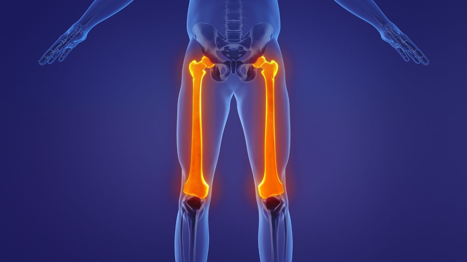
Osteoid osteoma can theoretically develop in any bone. However, in practice, it tends to prefer certain locations. The long and strong bones that bear the body’s weight are the most common sites.
Most frequently affected areas:
- Femur (thigh bone)
- Tibia (shin bone)
- Spine
- Humerus (upper arm bone)
- Wrist bones
- Metatarsal bones
- Heel bone
- Around the hip joint
In more than half of cases, the problem is located around the knee—either the distal femur or the proximal tibia. However, the exact location within the bone can significantly influence symptoms. When located in the cortical (outer hard shell) area of long bones, it may cause intense bone sclerosis and pain. If it is near a joint, symptoms may be more complex.
In joint-related areas such as the hip, patients may sometimes feel pain in the knee. This is called referred pain. Additionally, tumors located in the spine may cause painful scoliosis in children due to muscle spasm. Therefore, every child diagnosed with scoliosis and experiencing back pain should be carefully evaluated for this condition.
What Are the Diagnostic Steps and Imaging Methods?
Although the patient’s history and physical examination provide strong clues, advanced imaging methods are required for definitive diagnosis. It is common for patients to say, “My X-ray came back normal but my pain continues.” This is a situation frequently encountered.
Diagnostic methods include:
- Plain radiography
- Computed tomography (CT)
- Magnetic resonance imaging (MRI)
- Bone scintigraphy
X-ray is the first-step examination but is not always sufficient. In classical cases, bone thickening and a small central radiolucent area (nidus) can be observed. However, in joint-related locations or complex bone structures like hands and feet, X-rays may be inadequate and the tumor can be missed.
At this stage, computed tomography (CT) is considered the “gold standard.” CT shows bone structure in great detail and allows us to identify the exact location, size, and core of the tumor with millimetric precision. CT images are essential when planning treatment.
Magnetic resonance imaging (MRI) can sometimes be misleading. It often shows the surrounding edema excessively and may exaggerate the condition or cause confusion with bone infections (osteomyelitis) or malignant tumors. Therefore, while MRI is useful for evaluating soft tissues, CT is indispensable for detecting the nidus and making a definitive diagnosis.
Contact us for detailed information and an appointment!
Is It Confused With Growing Pains or Stress Fractures?
This is one of the most frequently asked questions by concerned parents. Unfortunately, the answer is yes; osteoid osteoma is often mistaken for other conditions, which can delay diagnosis. Many children are monitored for months or even years under the assumption of “growing pains.”
Conditions considered in differential diagnosis:
- Growing pains
- Stress fractures
- Bone infections
- Osteoblastoma
- Muscle strain
The difference between growing pains and osteoid osteoma is actually quite clear. Growing pains usually occur in both legs, often appear in the evening due to fatigue, and are relieved by massage. Osteoid osteoma pain, however, is located at a single point, is unilateral, does not improve with massage, and may worsen with touch. More importantly, growing pains do not wake the child from sleep, whereas osteoid osteoma does.
Stress fractures are typically seen in athletes or military personnel who perform high-intensity activities. In stress fractures, pain increases with activity (running, walking) and decreases with rest. In osteoid osteoma, the opposite occurs—the pain peaks during rest and at night. These distinctions are crucial for making the correct diagnosis.
Another important condition often confused with osteoid osteoma is “osteoblastoma.” Osteoblastoma is like the “big sibling” of osteoid osteoma. Although similar microscopically, osteoblastoma is larger than 2 cm and behaves more aggressively. It responds less to aspirin and requires surgical removal. In contrast, osteoid osteoma can be treated with simpler methods. CT imaging and lesion size are critical for making this distinction.
What Happens If It Is Not Treated, and Is Medication Alone Sufficient?
Osteoid osteoma is a self-limiting condition. In other words, even without surgical or interventional treatment, the tumor may eventually mature and fade over time (typically 3–7 years). Pain gradually decreases, and the lesion heals through calcification.
However, the main issue is the suffering the patient experiences during this long waiting period. Living with nightly pain for 5–6 years, being sleep-deprived, and constantly relying on high doses of painkillers is not acceptable for a child or active young adult.
Risks of long-term medication use include:
- Heartburn
- Stomach bleeding
- Stomach ulcers
- Kidney problems
- Bleeding disorders
- Hypertension
Therefore, treatment decisions should be based not on the biological behavior of the tumor but on the level of patient suffering. If pain cannot be controlled with medication, if the patient becomes tired of medication, experiences side effects, or if pain limits daily life, interventional treatment becomes necessary.
How Is Radiofrequency Ablation (RFA) Performed?
Radiofrequency ablation (RFA) is considered the gold standard treatment for osteoid osteoma today. Thanks to this technology, patients can avoid the risks and long recovery times associated with open surgery. The entire procedure is performed percutaneously, without any surgical incision.
Steps of the procedure:
- Anesthesia
- Localization with CT
- Needle insertion
- Thermal ablation
- Needle removal
The procedure is usually performed under general or spinal anesthesia so the patient feels no pain. The patient is placed on the CT table. Using CT images, the physician determines the exact location of the tumor with millimetric accuracy. Then, a special needle (electrode) is inserted through the skin to reach the nidus.
Once the needle tip reaches the center of the nidus, the radiofrequency generator is activated. The device creates heat at the needle tip through high-frequency vibrations. The tumor tissue is heated for approximately 4–6 minutes to temperatures up to 90°C. This high heat destroys the tumor cells and the nerve endings producing the pain (coagulation necrosis). After the procedure, the needle is removed, a small bandage is applied, and the process is completed.
Advantages of radiofrequency ablation:
- No incision
- No stitches required
- Same-day discharge
- Short procedure time
- Fast recovery
- Low complication risk
- High success rate
This method has a success rate of 90–95%. Most patients are surprised to notice that the severe pain they have endured for years disappears completely on the first night after treatment.
Contact us for detailed information and an appointment!
In Which Situations Are Surgery and Other Methods Preferred?
Although radiofrequency ablation (RFA) is the first choice, it may not be applicable or may be risky in some special situations. The heat generated during RFA spreads slightly from the needle tip into surrounding tissues. If the tumor is located in a sensitive area, this heat may damage nearby structures.
Situations requiring alternative approaches:
- Close proximity to nerves
- Close proximity to the skin
- Proximity to the spinal cord
- Proximity to the growth plate
- Suspicion of osteoblastoma
If the tumor is closer than 1.5 cm to a major nerve bundle, RFA may cause nerve damage or paralysis. In such cases, cryoablation (freezing) may be preferred. Cryoablation destroys the tumor by freezing it instead of heating it. Nerve tissue tolerates cold better than heat, and the ice ball formed during freezing is visible on imaging, allowing better control.
In growing children, growth plates (physes) are responsible for bone lengthening. If the tumor is very close to this plate, the heat generated by RFA could close the plate and stop bone growth, potentially causing length discrepancy or deformity. In such sensitive cases or when the tumor is located in a complex area, open surgery may be considered.
In open surgery, the tumor-bearing bone area is removed in a block (en-bloc excision) or curetted (scraped out). However, open surgery weakens the bone, increasing the risk of fracture and prolonging recovery. Therefore, it is used only when absolutely necessary. Even in modern surgical approaches, the goal is to perform the least invasive method possible with minimal incisions using CT guidance.
What Is the Recovery Process After Treatment and What Should Be Considered?
Recovery after radiofrequency or other minimally invasive treatments is generally very positive. Patients can usually stand up and walk a few hours after the procedure. Most are discharged the same day or the day after.
Expected conditions during recovery:
- Mild aching
- Tenderness at the entry site
- Rapid pain relief
- Return to daily activities
- Sleeping without medication
During the first week after the procedure, mild pain may occur at the needle entry site or within the bone, but this can be controlled with simple painkillers. Most patients immediately notice that their severe nighttime pain has completely disappeared.
However, there is one very important point: the disappearance of pain does not mean the bone immediately regains its full strength. The treated area remains biomechanically weak until full biological healing occurs. Especially in weight-bearing areas such as the femoral neck, the bone may be vulnerable during the recovery period.
Activity restrictions include:
- Avoiding high-impact sports
- No heavy lifting
- Avoiding running
- Avoiding contact sports
- Controlled walking
Generally, patients must avoid heavy sports or activities that strain the bone for the first 3 months. Low-impact exercises like walking or swimming are allowed, but activities such as football, basketball, or running should be postponed until complete remodeling occurs. Rarely, early excessive loading may lead to stress fractures or cracks in the treated area.
Finally, the possibility of recurrence should be addressed. Although success rates are high, literature reports recurrence rates between 10% and 20%. This risk is slightly higher in very young children. If recurrence occurs, the patient typically notices the return of pain months later. The good news is that a second RFA session usually provides nearly 100% success with complete resolution.
Frequently Asked Questions
In which age group and bones is osteoid osteoma most commonly seen?
This tumor generally appears in young individuals aged 10–30 and is most commonly observed in the femur and tibia. It settles in the cortical regions of long bones and is more frequently seen in males.
Why does osteoid osteoma cause increased nighttime pain?
Because tumor cells produce prostaglandins more intensely at night, pain becomes more severe. This type of pain temporarily improves with aspirin or NSAIDs.
What problems can osteoid osteoma cause in growing children?
If the tumor is close to the growth plate, it may cause limb length discrepancy, deformity, or gait abnormalities. Early diagnosis prevents these complications.
How does osteoid osteoma appear on X-ray or MRI?
X-rays show a small central lesion called a nidus and surrounding sclerotic bone tissue. MRI and especially CT provide a clearer view of the tumor and confirm the diagnosis.
Which bone conditions can be confused with osteoid osteoma?
Osteoblastoma, stress fracture, Brodie abscess, and some benign bone tumors may present similar findings. CT and biopsy (if needed) ensure accurate diagnosis.
Can osteoid osteoma be treated without surgery?
In some patients, if pain is manageable, medical monitoring may be considered. However, since this process can take years, surgical or radiofrequency ablation is preferred in most cases.
How is radiofrequency ablation applied in osteoid osteoma treatment?
Using CT guidance, a special needle is advanced into the nidus, and high-frequency energy is delivered to destroy the tumor. It is a minimally invasive method with a very short hospital stay.
How is osteoid osteoma surgically removed?
In surgical excision, the nidus is completely removed. Nowadays, minimal incision techniques are preferred. If the nidus is not fully removed, the pain may persist.
Does osteoid osteoma recur after treatment?
When performed correctly, the risk of recurrence is low. However, if the nidus is incompletely removed, the tumor may regrow and symptoms may return.
Does osteoid osteoma affect long-term bone health?
If untreated, it may cause chronic pain, movement limitation, muscle weakness, and bone deformities. With timely and proper treatment, bone health can be fully protected.
Blog Posts

Knee Replacement Surgery Prices
Ankara Ortopedi | Prof. Dr. Murat Demirel » Knee Treatments » Knee Replacement Surgery PricesThe [...]
18
Aug
Aug
Hip Replacement Surgery Prices
Ankara Ortopedi | Prof. Dr. Murat Demirel » Hip Treatments » Hip Replacement Surgery PricesThe [...]
18
Aug
Aug