

Ankle cartilage treatment includes all medical and surgical methods aimed at repairing damage to this vital tissue that covers the joint surface and ensures smooth movement. These injuries, usually occurring after trauma or repetitive strain, cause complaints such as deep pain that increases with activity, swelling, joint locking, and catching, which reduce quality of life. Treatment is personalized depending on the stage and size of the damage, ranging from activity modification, physical therapy, and biological injections such as PRP, to advanced surgical interventions like microfracture or cartilage transplantation.
| Disease Name | Ankle Cartilage Injury (Osteochondral Lesion) |
| Affected Area | Talus bone in the ankle joint |
| Main Causes | Trauma (especially sprains), repetitive microtraumas, circulatory disorders, sports injuries |
| Symptoms | Ankle pain, swelling, movement limitation, catching sensation, feeling of instability |
| Risk Factors | Sports activities, recurrent ankle sprains, anatomical deformities, obesity |
| Diagnostic Methods | Physical examination, magnetic resonance imaging (MRI), computed tomography (CT), X-ray |
| Treatment Methods | Conservative treatment (rest, load reduction, physical therapy), arthroscopic debridement, microfracture, mosaicplasty, autologous chondrocyte implantation |
| Surgical Necessity | Surgery may be required in symptomatic moderate-to-severe lesions unresponsive to conservative treatment |
| Need for Physical Therapy | Recommended post-surgery to restore joint mobility, strengthen muscles, and regulate loading |
| Possible Complications | Insufficient cartilage healing, persistent pain, joint stiffness, recurrence |
| Recovery Period | May vary between 6–12 weeks depending on lesion size and applied treatment |
| Need for Follow-up | Clinical and imaging follow-up recommended to monitor healing |


Prof. Dr. Murat Demirel
Orthopedics and Traumatology Specialist
Orthopedics Specialist Prof. Dr. Murat Demirel was born in Ankara in 1974. He completed his primary education at Ankara Kavaklıdere Primary School and his secondary and high school education at Ankara Atatürk Anatolian High School. Dr. Demirel graduated from Ankara University Faculty of Medicine in 1998 and completed his residency in Orthopedics and Traumatology at Ankara Numune Training and Research Hospital, 1st Orthopedics and Traumatology Clinic, in 2004.
PhD
Ankara University Institute of Health Sciences
Specialization
Ankara Numune Training and Research Hospital, 1st Orthopedics Clinic
Medical School
Ankara University Faculty of Medicine
Yazı İçeriği
Why Is Ankle Cartilage Tissue So Important for Our Body?
The ankle joint is essentially a pulley system formed by the union of three bones (tibia, fibula, and talus). For this system to function smoothly, the cartilage covering the joint surfaces of the bones has two vital roles. First, it prevents bones from rubbing against each other during joint movements, creating an incredibly low friction coefficient. This allows the joint to move fluidly and painlessly with almost no energy loss. Its second important function is acting as a shock absorber. When we walk, several times our body weight loads onto the ankle, and while running, even more. Thanks to its flexible structure, cartilage tissue absorbs these loads and prevents damage to the bones.
Perhaps the most critical feature of this tissue, which makes it so unique, is that it has almost no self-healing ability. The reason is that, unlike our skin or muscles, it is not nourished by blood vessels. Cartilage receives nutrients from a special fluid inside the joint called “synovial fluid,” aided by the pumping effect created by pressure changes during movement. Not having a blood vessel network means that when an injury occurs, healing cells and repair factors cannot reach the damaged area. That is why when cartilage damage occurs, instead of waiting for it to heal on its own, it becomes necessary to adopt an active treatment approach to solve this problem.
What Are the Causes Behind Ankle Cartilage Damage?
Many people think that cartilage problems are only related to aging, but in the ankle, the situation is quite different. Unlike the knee joint, most ankle cartilage injuries occur as a result of previous trauma rather than age-related wear and tear. Even an “ordinary sprain” that is underestimated may actually be the beginning of serious and permanent damage to the cartilage surface. The main risk factors and causes leading to cartilage damage are:
- Sudden trauma (especially ankle sprains)
- Repetitive microtraumas
- Chronic joint laxity (instability)
- Joint alignment problems
- Previous joint fractures
- Obesity
- Certain rheumatic diseases
Among these listed factors, the most common scenario is ankle sprains. During a sprain, the talus bone hits the edge of the tibia, causing a bruise or fracture in both the cartilage and the underlying bone. We call this condition “osteochondral lesion” (OCL). Most patients expect the pain to pass after the sprain, but deep aching that continues for weeks or even months can be a sign of underlying cartilage damage. Similarly, joint laxity (instability) that develops after recurrent sprains causes abnormal joint movements with each step and gradually wears down the cartilage like sandpaper. Therefore, in unresolved or recurrent ankle problems, cartilage injury should be the first consideration.
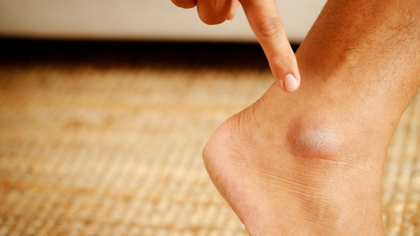
What Are the Typical Symptoms of Ankle Cartilage Damage?
The symptoms of ankle cartilage damage may be mild and vague at first, but as the damage progresses over time, they become more noticeable. The most common complaints that make patients consult a doctor are usually a sense that “something is wrong” in the joint. If you are experiencing one or more of the symptoms below, you may have an underlying cartilage problem.
The most common symptoms of ankle cartilage damage are:
- Deep aching pain
- Swelling that increases with activity
- Catching sensation in the joint
- Locking
- “Click” or “pop” sounds
- Movement restriction
- Feeling of instability or looseness
Pain is usually the main symptom, and patients often describe it as “something stabbing deep inside my ankle” or “my bones are aching.” This pain decreases at rest but worsens with weight-bearing or prolonged standing. Swelling typically occurs after activity and represents the body’s inflammatory response to a problem inside the joint.
The most important and diagnostic symptoms are the “mechanical symptoms,” which include catching, locking, and noises. Catching is the sensation of a brief obstruction during movement. Locking is more serious; the joint becomes stuck in a certain position, and the patient cannot move it without making a small maneuver. These symptoms usually indicate that a damaged cartilage or bone fragment has broken off and turned into a free body inside the joint (“joint mouse”). This free fragment gets trapped during joint movements, both causing these sensations and scratching the healthy cartilage surfaces, worsening the damage. Therefore, the presence of mechanical symptoms often indicates the need for more urgent treatment.
Contact us for detailed information and an appointment!
How Does a Doctor Diagnose Ankle Cartilage Damage?
The first and most important step in correct treatment is an accurate diagnosis. Diagnosing cartilage damage is a three-stage process that requires gathering clues like a detective.
Listening to the Patient’s History (Anamnesis)
This process begins with a detailed interview. Details such as when and how your complaints started, the mechanism of the first injury, the character of the pain (when it increases, when it decreases), whether you experience catching or locking, and previous treatments provide the first diagnostic clues.
Detailed Physical Examination
Next comes a careful physical examination of your ankle. Your walking pattern is observed, and the joint is checked for swelling, deformity, or tenderness. The range of motion of your ankle (upward-downward, inward-outward movements) is assessed both actively and passively. Special tests are applied to detect possible ligament laxity. The location of tenderness during examination offers valuable information about the possible site of cartilage damage.
Imaging Methods
If physical examination findings suggest cartilage damage, imaging methods are used to confirm the diagnosis and clearly assess the extent of the damage.
X-ray: Usually the first test requested. It can show fractures, bone protrusions, or advanced osteoarthritis. However, since cartilage is soft tissue, it cannot be seen on X-rays. Therefore, a “clean” X-ray in a patient with ankle pain does not necessarily mean the cartilage is healthy.
Magnetic Resonance Imaging (MRI): Considered the “gold standard” in diagnosing ankle cartilage injuries. MRI shows us the inside of the joint almost three-dimensionally. It reveals cartilage thickness, surface irregularities, the presence of detached fragments, edema (bone marrow edema) or cysts in the bone under the cartilage. It is the most important roadmap directly affecting the treatment method (non-surgical, microfracture, cartilage transplantation, etc.).
Computed Tomography (CT): May be required to evaluate bone structure in detail, see cyst boundaries, or plan after complex fractures. In complex cases, both MRI and CT may be used together to determine the most accurate surgical strategy.
Are There Treatment Options for Ankle Cartilage Injury Before Surgery?
Absolutely yes. Not every ankle cartilage injury means immediate surgery. Especially in early-stage, small-sized, stable (non-displaced), and non-mechanical (no catching or locking) lesions, our primary goal is to relieve the patient’s symptoms with non-surgical methods. The main aim of these treatments is not to completely regenerate damaged cartilage, but to control pain and inflammation, improve joint function, and slow disease progression.
The main conservative treatment methods applied initially are:
- Activity modification
- Ice application
- Pain relievers and anti-inflammatory medications
- Physical therapy and exercise
- Ankle braces or bandaging
- Weight control
Among these methods, physical therapy plays a key role in long-term success. The goal of physical therapy is not only simple exercises but also strengthening the muscles around the ankle (especially the calf and anterior leg muscles), and improving balance sense (proprioception) to reduce joint load and support the joint like a natural brace. Strong and balanced muscles help reduce stress on damaged cartilage, relieve pain, and prevent possible future injuries.
What Injections Are Used in Ankle Cartilage Problems?
When physical therapy and general measures are not sufficient, intra-articular injection therapies may be an option to control symptoms more effectively. These injections aim to improve the biological environment inside the joint and reduce pain rather than directly repairing cartilage.
Corticosteroid (Cortisone) Injections: When injected into the joint, they strongly suppress local inflammation and swelling, providing rapid pain relief. However, this effect is temporary (lasting a few weeks to months), and since cortisone itself can damage cartilage if used frequently, its use is limited. It is generally considered a “bridge therapy” to suppress severe pain flare-ups or provide relief before surgery.
Hyaluronic Acid Injections (Viscosupplementation): Synthetic forms of hyaluronic acid, one of the main components of healthy joint fluid, are injected into the joint. Also known as “rooster comb injection” or “joint fluid supplement.” The aim is to increase joint lubrication, reduce friction, and act as a mild shock absorber to relieve pain. Though less common in ankles compared to knees, it may benefit some patients in early-stage ankle arthritis.
Platelet-Rich Plasma (PRP) Injections: In this method, a small amount of the patient’s blood is drawn and centrifuged to obtain plasma rich in platelets (healing cells). Platelets contain many growth factors that initiate tissue repair. Injecting this concentrated plasma into the damaged cartilage area or joint aims to trigger the body’s natural healing mechanisms and accelerate repair.
Stem Cell / Bone Marrow Aspirate Concentrate (BMAC) Injections: One of the most advanced applications of regenerative medicine. Bone marrow is usually drawn from the pelvic bone under local anesthesia and processed to concentrate mesenchymal stem cells and repair factors. Stem cells are “master” cells capable of differentiating into various tissues, including cartilage. Injecting this concentrate into the joint aims to initiate cartilage regeneration at the damaged site. Although promising, research is still ongoing regarding their effectiveness and best patient selection.
What ‘Cartilage Transplantation’ Techniques Are Used for Large Ankle Cartilage Injuries?
If cartilage damage is large, if there are serious underlying bone problems such as cysts, or if methods like microfracture fail, more comprehensive “restorative” surgeries that aim to replace the damaged area with new, healthy cartilage tissue come into play. These methods are essentially based on performing a “cartilage transplant.”
Osteochondral Autograft Transfer (OATS / Mosaicplasty): This method can be compared to moving healthy sod patches from another part of your garden to a thinning area. During surgery, cylindrical bone-cartilage plugs are harvested from a non-weight-bearing area of the patient’s knee joint. These plugs are transplanted into the damaged ankle area side by side, like a mosaic pattern. Its biggest advantage is replacing the damaged area with living, original hyaline cartilage, which is the highest quality biologically. It offers long-term, permanent results especially in young and active patients. Its disadvantage is possible discomfort at the donor knee site.
Osteochondral Allograft Transplantation (Donor Transplant): Preferred when cartilage damage is very large and sufficient autografts cannot be harvested from the patient’s knee. In this method, fresh bone-cartilage tissue (allograft) taken from a deceased donor, obtained from special tissue banks, is transplanted into the damaged area. This avoids a second surgery on the patient and allows repair of large areas in one piece. Disadvantages include difficulty in finding suitable donor tissue and high cost.
Autologous Chondrocyte Implantation / Matrix-Associated Chondrocyte Implantation (ACI/MACI): A two-stage, high-tech “cell seeding” technique. In the first surgery (usually minimally invasive), a small sample of healthy cartilage is taken from the patient. This sample is sent to a special laboratory where chondrocytes (cartilage cells) are isolated and cultured for 3–6 weeks to produce millions of cells. In the second surgery, these new cells are implanted into the damaged area on a special membrane (matrix) or under a patch. The goal is for the body to use these implanted cells to gradually form new, healthy cartilage tissue. It is considered an option especially in very large cartilage losses where no other method is suitable.
Contact us for detailed information and an appointment!
Frequently Asked Questions
What is ankle cartilage injury and how does it occur?
Ankle cartilage injury is the damage of the cartilage tissue covering the ankle joint that prevents the bones from rubbing against each other. It usually occurs due to ankle sprains, repetitive trauma, sports injuries, or circulatory disorders in the joint. Sometimes cartilage injury may also occur together with small fractures.
What are the symptoms of ankle cartilage injury?
Pain in the ankle, swelling, a catching or locking sensation during movement, sometimes mild restriction of movement, and occasional clicking or grinding sounds from the joint may occur. Pain usually increases with weight-bearing or prolonged standing.
How is ankle cartilage injury diagnosed?
Patient history and physical examination findings are important in diagnosis. X-rays usually show bone structure but are not sufficient to detect cartilage damage. Magnetic resonance imaging (MRI) is most often used for a definitive diagnosis.
Can cartilage injury heal on its own?
Because cartilage tissue lacks blood vessels, its ability to heal on its own is very limited. In mild injuries, symptoms may temporarily decrease, but complete healing is generally not possible. If the injury progresses, osteoarthritis and permanent problems may develop in the joint.
How is ankle cartilage injury treated?
Treatment depends on the extent of the injury and the patient’s symptoms. In mild cases, rest, cold application, pain relievers, and physical therapy may be sufficient. In more severe cases, PRP, stem cell, hyaluronic acid injections, or arthroscopic surgery may be considered.
When is surgery necessary?
In large, deep, or recurrent cartilage injuries, and in patients whose complaints persist despite conservative treatment, arthroscopic surgery may be required. Cartilage repair, microfracture, or mosaicplasty may be performed.
Is full recovery possible after cartilage injury?
Although cartilage tissue does not return completely to its original state, with early diagnosis and appropriate treatment, complaints can be significantly reduced and joint function preserved. However, in advanced cases, the risk of permanent restriction and pain increases.
Is return to sports possible after ankle cartilage injury?
After appropriate treatment and rehabilitation, most patients can return to light sports. However, for high-intensity and strenuous sports, doctor approval is essential and the return-to-sport process must be carefully planned.
What can be done to prevent cartilage injury?
Strengthening the ankle muscles, wearing appropriate footwear, paying attention to ground conditions, and not neglecting warm-up exercises in sports help prevent cartilage injuries. Avoiding repetitive sprains is also important.
What happens if ankle cartilage injury progresses?
Untreated cartilage injuries may lead to osteoarthritis, chronic pain, and limited mobility over time. Therefore, early diagnosis and treatment are very important.
Blog Posts

Knee Replacement Surgery Prices
Ankara Ortopedi | Prof. Dr. Murat Demirel » Knee Treatments » Knee Replacement Surgery PricesThe [...]
18
Aug
Aug
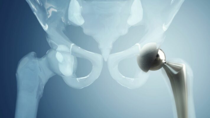
Hip Replacement Surgery Prices
Ankara Ortopedi | Prof. Dr. Murat Demirel » Hip Treatments » Hip Replacement Surgery PricesThe [...]
18
Aug
Aug