

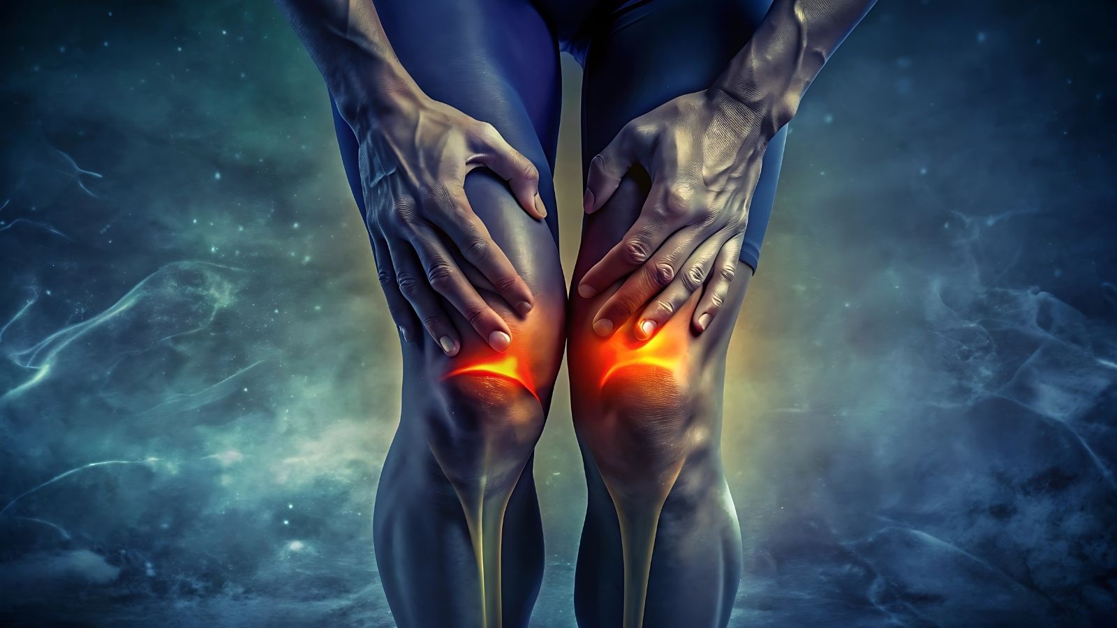
An osteochondroma is a benign bone–cartilage tumor that develops outward from the bone surface and is most often detected during childhood and adolescence. Although it is asymptomatic in most cases, it has growth potential and therefore requires regular clinical follow-up.
The symptoms of osteochondroma appear as a palpable hard prominence in the mass region, pain developing over time, and restriction of movement due to mechanical irritation. When pressure on nearby tissues develops, nerve compression or vascular involvement may also be observed.
Diagnostic methods for osteochondroma are based on demonstrating continuity with the underlying bone on plain radiographs. MRI and CT scans are important for assessing the thickness of the cartilage cap, its relationship with surrounding tissues, and the risk of malignant transformation.
Treatment of osteochondroma is performed with surgical excision in lesions that cause symptoms, grow rapidly, or affect neurovascular structures. After complete excision, the risk of recurrence is low, and the operation safely supports functional recovery.

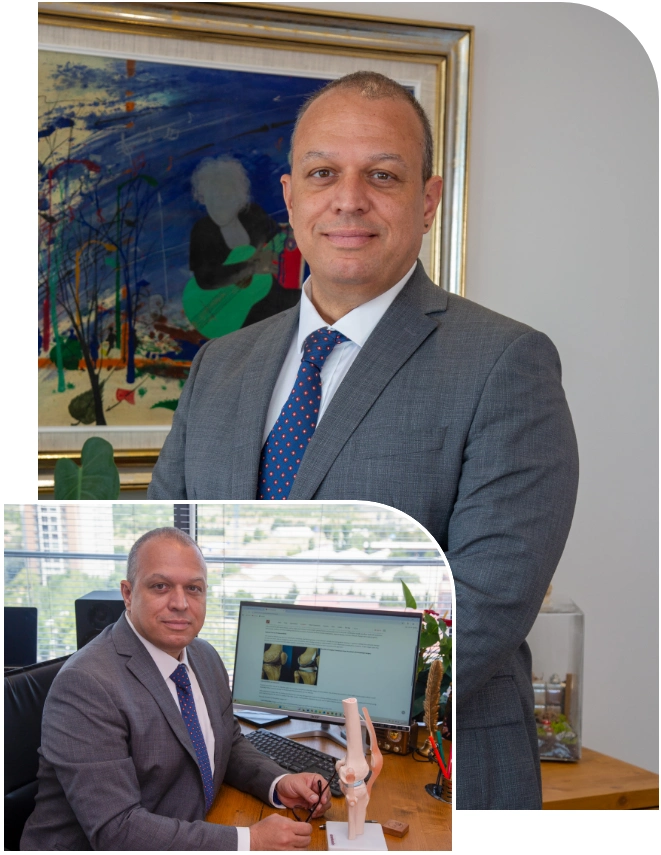
Prof. Dr. Murat Demirel
Orthopedics and Traumatology Specialist
Orthopedics Specialist Prof. Dr. Murat Demirel was born in Ankara in 1974. He completed his primary education at Ankara Kavaklıdere Primary School and his secondary and high school education at Ankara Atatürk Anatolian High School. Dr. Demirel graduated from Ankara University Faculty of Medicine in 1998 and completed his residency in Orthopedics and Traumatology at Ankara Numune Training and Research Hospital, 1st Orthopedics and Traumatology Clinic, in 2004.
PhD
Ankara University Institute of Health Sciences
Specialization
Ankara Numune Training and Research Hospital, 1st Orthopedics Clinic
Medical School
Ankara University Faculty of Medicine
Yazı İçeriği
What Is an Osteochondroma?
An osteochondroma is a benign (noncancerous) bone tumor that develops on the bone and usually appears near the growth plates. It is typically detected during adolescence and is commonly seen at the ends of long bones, especially around the knee. If it does not cause pain, it does not require treatment; however, if it grows and puts pressure on surrounding tissues, it can be surgically removed. It rarely transforms into a malignant tumor.
How Does an Osteochondroma Form?
In childhood, at the ends of our long bones there are very special production centers that we call the “growth plate” or “physis.” This is like a factory where cartilage cells continuously divide and proliferate, later turning into bone and allowing our height to increase. This factory operates at an incredible speed.
An osteochondroma is essentially the story of some cells that escape from this rapidly working factory. Some cartilage cells in the growth plate deviate from their normal vertical growth path and start to grow sideways, toward the side of the bone. You can think of this like an unexpected branch growing sideways from the trunk of a tree. This side branch has the same structure as the main bone; it contains bone marrow inside and has a hard bony shell on the outside. However, on its top, it carries a “cap” of cartilage that retains growth potential.
This cartilage cap is very important to us because it is the area that enables the tumor to grow. As your child’s height increases and the growth plates remain active, this cartilage cap also stays active and the protrusion continues to grow. When puberty ends and the growth plates close, theoretically the growth of this protrusion also stops. In other words, this condition is part of a dynamic and living process.
What Are the Symptoms of an Osteochondroma?
The vast majority of our patients actually have no complaints. Most of the time, we detect these protrusions incidentally on X-rays taken for another reason, such as a suspected sprain. However, when they become prominent or are located in strategic areas, they begin to give certain signals.
The most common symptoms of osteochondroma are:
- Hard swelling
- Painless mass
- Restriction of movement
- Sensation of mechanical catching
- Localized tenderness
- Muscle weakness
- Numbness or tingling
- Color change due to vascular compression
- Limb deformity
- Short stature
- Cosmetic concerns
The most frequent finding among these is a palpable, fixed mass. Since the mass is attached to the bone, it does not move side to side under the skin; it remains fixed. Pain usually does not arise from the tumor itself but from the muscles or tendons that rub over the tumor.
In Whom and in Which Areas Does This Condition Occur?
This is a problem of “growing bones.” Therefore, the majority of our patients are children and adolescents. We typically make the diagnosis in young people between the ages of 10 and 20. The incidence is similar in girls and boys, although we see it slightly more often in boys.
As for its location in the body, these lesions prefer the most mobile and fastest growing regions. The area around the knee is where bone growth is fastest in our body. Therefore, we most commonly see osteochondromas at the lower end of the femur (thigh bone) or the upper end of the tibia (shin bone). The second most common location is the upper end of the humerus (arm bone), that is, the shoulder region. However, they can also appear in the pelvis, scapula, ribs, and even in the bones of the hands and feet.
They are very rarely seen in the facial bones because the growth mechanism of facial bones is different from that of long bones. If there is a single protrusion in a single bone, we call this a “solitary osteochondroma,” and about 85% of cases fall into this group.
What Is Hereditary Multiple Exostoses (HME)?
The remaining 15% of cases fall into a group that requires more attention and care from us. If a child has multiple protrusions in several bones, we call this “Hereditary Multiple Exostoses” or HME for short. This condition has a genetic basis. It occurs due to functional abnormalities in genes called EXT1 or EXT2, which are inherited from the mother or father.
In children with HME, the issue is not simply a single bony lump. The growth potential of the bones can also be affected in these children. Bones may not reach their expected length or may grow in a curved manner. In particular, mismatch in the growth of the two forearm bones (radius and ulna) can cause deformities at the wrist or elbow dislocations. Therefore, the follow-up of a child diagnosed with HME must be planned not only based on current complaints, but also with regard to future skeletal health and postural deformities.
Contact us for detailed information and an appointment!
How Is the Diagnosis Made?
The diagnostic process actually begins with the physical examination we perform in the clinic. However, to understand the bony structures that we cannot see with our eyes, we need radiological imaging.
The methods we use at the diagnostic stage are:
- X-ray (Plain radiograph)
- Computed Tomography (CT)
- Magnetic Resonance Imaging (MRI)
In almost all cases, a simple X-ray is sufficient for diagnosis. On the radiograph, we see the characteristic “cauliflower-like” or “pedunculated” structure extending outward from the bone. Demonstrating that the interior of this protrusion is continuous with the main bone is enough to confirm the diagnosis.
However, in some cases, X-rays are not sufficient. We may want to obtain a three-dimensional map with Computed Tomography (CT), especially in complex regions such as the pelvis or spine.
Our most critical investigation is Magnetic Resonance Imaging (MRI). We do not request MRI for every patient. However, if we are going to make a surgical decision or suspect malignant transformation, MRI is essential. This is because on an X-ray we can see the bone, but we cannot see the famous “cartilage cap” on top of the tumor. MRI is the method that shows the thickness and structure of this cartilage cap most clearly. We also use this test to understand how close the tumor is to surrounding vessels and nerves.
Is There a Cancer Risk?
This is the issue that families fear the most, are often hesitant to ask about, but absolutely need to know. Let me start with reassurance: the risk of solitary osteochondromas turning into cancer (chondrosarcoma) is extremely low. Statistically, this rate is below 1%. In other words, in ninety-nine out of one hundred patients, these masses remain benign throughout life. In patients with HME who have multiple lesions, this risk is somewhat higher, but still not very common.
We physicians have certain red flags that make us wonder, “Could there be a malignant change?” The most important of these is the thickness of the cartilage cap.
Situations that raise suspicion of malignant transformation include:
- Growth in adulthood
- Cartilage cap thicker than 2 cm
- Sudden onset of pain
- Irregular borders
- Formation of a soft tissue mass
Since growth is continuing in children, a cartilage cap up to 3 cm thick can be considered normal. However, in an adult whose skeletal development is complete, the cartilage cap should be very thin, almost like a sheet of paper. If in an adult patient we measure a cartilage cap thicker than 2 cm on MRI, this raises an alarm and leads us toward biopsy or surgical planning. In addition, if a mass that has been silent for years suddenly starts to grow or become painful in the patient’s 40s or 50s, we must take this seriously.
What Is the Follow-Up Process Like?
If an osteochondroma is detected in your child and there is no pain or restriction of movement, the best treatment is “to do nothing.” We call this “active surveillance” in medical terms.
Active surveillance does not mean abandoning the patient to their fate. We periodically (usually every 6 or 12 months) take X-rays to monitor the tumor’s growth rate and shape. Our goal is to avoid unnecessary surgery. Because every surgical intervention—especially those performed near the growth plate—carries a risk of stopping or disrupting bone growth.
Nature has a wonderful balance. As the child grows, the muscles and tendons generally adapt to pass around this protrusion. Therefore, rather than rushing into an early, hasty surgery, the safest approach is to wait until the bone matures. Once skeletal maturity is reached and the growth plates close, the tumor’s growth also stops, so we reduce the frequency of follow-up or discontinue it altogether.
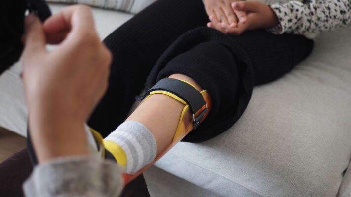
What Are the Non-Surgical Treatment Options?
Sometimes the mass may cause pain but not to an extent that warrants surgery. In these “intermediate periods,” we use non-surgical methods to provide patient comfort. Remember, no medication or cream will eliminate this bony protrusion; they only reduce the symptoms it causes.
The non-surgical methods we use include:
- Pain-relieving medications
- Cold application (Ice)
- Activity modification
- Physical therapy
- Stretching exercises
- Muscle strengthening
- Special pads or supports
- Shoe modifications
For example, if a protrusion on the tibia rubs against the edge of the shoe while running, placing a soft pad in that area of the shoe or changing the shoe model may solve the problem. If a mass around the knee is straining the tendons, stretching those tendons with physical therapy may reduce friction and pain. Our aim here is to make the patient’s daily life manageable without surgery.
Contact us for detailed information and an appointment!
When Is Surgery Necessary?
If problems persist despite observation and supportive treatment, or the location of the tumor creates risk, it may be time to proceed to surgery. When making a surgical decision, we carefully weigh the risk–benefit balance.
Situations that require surgical intervention include:
- Severe pain
- Progressive growth
- Restriction of joint movement
- Nerve compression
- Vascular compression
- Tendon impingement
- Severe deformity
- Fracture formation
- Bursitis
- Suspicion of malignancy
- Cosmetic deformities
If the tumor is compressing major vessels or nerves behind the leg, we cannot afford to wait. Because such compression can lead to irreversible damage (such as paralysis or aneurysms). In addition, if bone deformities in HME patients impair the child’s ability to walk or use their hand, we plan surgery not only to remove the tumor but also to correct the bone alignment.
How Is Surgical Treatment Performed?
The method we use in osteochondroma surgery is called “marginal excision.” This term may sound technical, but the logic is simple: remove the tumor from its root.
In surgery, it is not enough to just shave down the visible bump. If we do not completely remove the cartilage cap and the surrounding sheath, the tumor may regrow from the microscopic cells left behind. This is very similar to cleaning weeds in a garden; if you only cut off the leaves, they will grow back, but if you remove the root completely from the soil, they will not.
During surgery, we go down to the base of the tumor where it attaches to the main bone. Using a chisel or a power saw, we separate the tumor from the main bone. Our greatest care at this stage is to protect the nerves and vessels passing around or beneath the tumor. Sometimes large tumors may displace or push these delicate structures. Therefore, surgical planning is done with great precision. In cases where the tumor is adherent to major vessels, we may perform the operation as a team with cardiovascular surgeons to ensure a safe procedure.
What Is the Recovery Process Like?
The postoperative course usually progresses faster than you might expect. In most patients, healing is rapid because we do not interfere with the main load-bearing body of the bone, but only remove the excess on top.
Important stages in the recovery period include:
- Length of hospital stay
- Wound care
- Removal of stitches
- Permission for weight-bearing
- Range-of-motion exercises
- Control of swelling (edema)
- Scar massage
Generally, our patients are discharged the day after surgery. During the first two weeks, we restrict contact between the wound and water to allow healing and closure of the stitches. Medications are usually sufficient for pain control during this period.
If surgery is performed on the leg, we allow the patient to stand up and walk to the bathroom using crutches as early as the next day. The time to bear full weight and abandon crutches varies between 2 and 4 weeks depending on the extent of surgery. We begin movements of the knee or shoulder joint immediately, because leaving a joint immobile for too long can lead to stiffness.
When Can Patients Return to Sports?
This question is particularly critical for young athletes and active individuals. Our usual answer to “Doctor, when can I return to the field?” is 3 to 4 months.
Why do we wait this long? Because when we separate the tumor from the bone, a weakened area may remain in the main bone. The bone’s repair of this area and restoration of its former strength is a biological process that cannot be accelerated. If patients return to high-impact sports such as running, jumping, or contact sports too early before the bone is fully healed, stress fractures or cracks can develop at that weak point.
During this 3–4-month period, the patient does not remain inactive; they maintain conditioning with non–weight-bearing activities such as swimming or stationary cycling. Once we see on X-rays that the bone has completely healed, we give the “you can return to the field” approval.
What Are the Long-Term Outcomes?
In solitary osteochondromas, the risk of recurrence after successful surgery is between 2% and 5%, which is quite low. In general, the patient leaves this problem behind completely and returns to normal life.
However, in patients with HME, the situation is a bit different. New protrusions may form in other parts of the body, or existing ones may grow. Therefore, we continue to follow these patients into adulthood, especially monitoring regions such as the pelvis and scapula, which are easily overlooked.
Frequently Asked Questions
In which age group and bones is osteochondroma most commonly seen?
Osteochondroma usually develops during childhood and adolescence. It is most commonly seen around the growth plates of long bones such as the femur, tibia, and humerus. Its growth ceases once skeletal maturity is reached.
Under what circumstances is growth of an osteochondroma dangerous?
If an osteochondroma continues to grow after adulthood, the risk of malignant transformation (chondrosarcoma) should be considered. Sudden growth, pain, or signs of nerve compression increase this likelihood.
What problems can an osteochondroma cause apart from bone pain?
It can cause pressure on surrounding muscles, nerves, or blood vessels. This may lead to numbness, restricted movement, circulatory impairment, or cosmetic problems.
Can osteochondroma be hereditary?
Yes, in a hereditary condition called multiple hereditary exostoses, numerous osteochondromas form throughout the body. These patients have a higher risk of deformity and malignant transformation.
How is osteochondroma distinguished on X-ray?
On X-ray, a stalked or broad-based bony protrusion covered with cartilage extending outward from the bone is seen. CT and MRI allow a more detailed evaluation of the thickness of the cartilage cap and its relationship with surrounding tissues.
In which osteochondroma cases is surgery mandatory?
Osteochondromas that cause pain, restriction of movement, vascular or nerve compression, cosmetic deformity, or raise suspicion of malignancy must be surgically removed.
How is osteochondroma surgery performed and how long does it take?
Surgery is performed by completely removing the tumor together with its bone and cartilage components. In small and superficial lesions, the procedure is short and hospital stay is usually brief.
Is there a risk of recurrence after osteochondroma surgery?
Osteochondromas that are completely removed rarely recur. However, in incompletely excised lesions or hereditary cases, the risk of new formation is higher.
What complications may develop if osteochondroma is not treated?
Over time, it may grow and lead to nerve and vessel compression, bone deformities, or rarely malignant transformation. Therefore, regular follow-up is important.
What is the recovery process like after osteochondroma treatment?
After surgery for small and superficial osteochondromas, patients can return to daily activities within a few days. In deeper or larger lesions, the healing and rehabilitation process may take several weeks.
Blog Posts
What Is a Tendon Rupture? Symptoms, Causes, and Treatment Methods
Ankara Ortopedi | Prof. Dr. Murat Demirel » General » What Is a Tendon Rupture? [...]
13
Apr
Apr
What Is an Orthosis? Why Is an Orthosis Used?
Ankara Ortopedi | Prof. Dr. Murat Demirel » General » What Is an Orthosis? Why [...]
13
Apr
Apr

Knee Replacement Surgery Prices
Ankara Ortopedi | Prof. Dr. Murat Demirel » Knee Treatments » Knee Replacement Surgery PricesThe [...]
18
Aug
Aug
Hip Replacement Surgery Prices
Ankara Ortopedi | Prof. Dr. Murat Demirel » Hip Treatments » Hip Replacement Surgery PricesThe [...]
18
Aug
Aug