

A lipoma is a benign soft tissue tumor that forms through the benign proliferation of subcutaneous fat tissue. It usually grows slowly and has a painless course. Diagnosis is most often made by clinical evaluation, and the risk of malignancy is quite low.
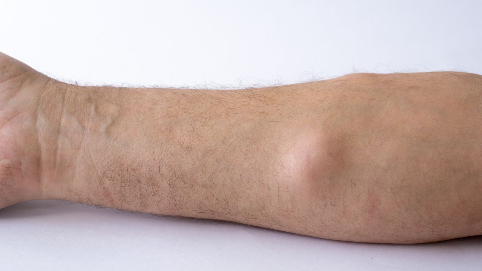
The symptoms of a lipoma present as a soft, mobile mass with clearly defined borders under the skin. When it reaches larger sizes, it may cause mild pain due to cosmetic discomfort or pressure on surrounding tissues.
Diagnostic methods for lipoma are based on confirming that the lesion originates from fat tissue with ultrasonography in addition to physical examination. When necessary, MRI is used to further detail the mass boundaries and its relationship with deeper tissues.
Treatment of lipoma is performed with surgical excision in cases that are symptomatic, rapidly growing, or causing cosmetic problems. The recurrence rate after surgery is low, and when complete excision is achieved, the need for long-term follow-up is minimal.


Prof. Dr. Murat Demirel
Orthopedics and Traumatology Specialist
Orthopedics Specialist Prof. Dr. Murat Demirel was born in Ankara in 1974. He completed his primary education at Ankara Kavaklıdere Primary School and his secondary and high school education at Ankara Atatürk Anatolian High School. Dr. Demirel graduated from Ankara University Faculty of Medicine in 1998 and completed his residency in Orthopedics and Traumatology at Ankara Numune Training and Research Hospital, 1st Orthopedics and Traumatology Clinic, in 2004.
PhD
Ankara University Institute of Health Sciences
Specialization
Ankara Numune Training and Research Hospital, 1st Orthopedics Clinic
Medical School
Ankara University Faculty of Medicine
Yazı İçeriği
What Is a Lipoma?
A lipoma is a slowly growing, usually harmless, soft fatty tissue tumor under the skin, medically referred to as a “lipoma.” It is commonly seen on areas such as the back, shoulders, arms, or neck. It is usually painless and does not pose a serious health risk. However, when it grows and causes discomfort or cosmetic concern, it can be removed surgically.
How Does a Lipoma Form in Our Body?
Among the lumps we occasionally feel in our body, lipomas are the most common. Technically, they fall into the “mesenchymal tumor” class, but this term should not worry you. Lipomas occur through the uncontrolled yet benign proliferation of mature fat cells (adipocytes) in the body. When you examine a lipoma by touch, you usually feel a soft, slippery mass just beneath the skin, like a rubbery ball. This mobility is one of the most characteristic features of a lipoma; when you press on it, it may shift to the sides.
From a pathological perspective, we see a thin, fibrous capsule surrounding a typical lipoma. This capsule holds the fatty tissue together, like a sheath, and clearly separates the mass from the surrounding tissues. This structure is an advantage during surgery because the mass can be peeled away from its surroundings like a fruit being separated from its peel. The internal content of the lipoma is identical to the yellow fat tissue in other parts of the body; the difference is that the fat cells are clustered together in one lump.
While superficial lipomas are often considered a cosmetic problem, the main group we focus on in orthopedic practice are the “deep-seated” ones. These are not located just under the skin but beneath the fascia (the sheath around muscles), between muscles, or within the muscle tissue itself. A deep lipoma may feel more firm and fixed compared to surface lipomas. Masses in these areas can become more than just a lump; they have the potential to compress nearby anatomical structures. Therefore, the management of a deep lipoma requires a much more complex and meticulous approach than a simple subcutaneous lump.
Why Do Lipomas Appear and What Are the Triggering Factors?
One of the most common questions our patients ask in the clinic is why these lumps form and whether they have done something wrong to cause them. Scientifically, the exact mechanism behind lipoma formation is not yet 100% clear. However, available data show that this condition does not arise from a single cause but rather from a combination of multiple factors. Contrary to common belief, there is no clear and direct evidence linking dietary habits to lipoma formation.
The strongest determining factor is genetic predisposition. Comprehensive genetic analyses have shown chromosomal abnormalities in approximately two-thirds of lipoma cases. In particular, rearrangements in specific regions of chromosome 12 and the activity of a gene called HMGA2 play a key role in the accumulation of fat cells in this way. Therefore, if you have a family history of multiple lipomas in your parents or close relatives, your likelihood of having a genetic predisposition is high.
In addition, physical trauma is thought to be a possible trigger. A strong blow to a particular area may disrupt the local tissue healing process or create chronic irritation, laying the groundwork for fat tissue accumulation. In this process, called “metaplasia,” damaged tissue can transform into fat tissue. However, a lipoma does not form at every site of trauma; this is more commonly seen in individuals with a genetic predisposition. Obesity or significant weight gain can cause existing lipomas to enlarge, but they are not the sole direct cause of lipoma formation. The fact that lipomas do not disappear with weight loss also indicates that this tissue has a different metabolism than normal body fat.
Factors thought to play a role in lipoma formation include:
- Genetic predisposition
- Chromosomal abnormalities
- HMGA2 gene
- Physical trauma
- Chronic irritation
- Metaplasia process
- Advanced age
- Obesity
What Are the Symptoms of a Lipoma and How Does It Feel?
A typical lipoma usually develops silently. Most patients notice it when they feel a lump while showering or getting dressed. Standard lipomas are painless, soft, and do not cause any discoloration or sores on the overlying skin. However, as the lipoma’s depth of location increases or its size grows, the complaints it causes can vary. Deep lipomas, which are especially important from an orthopedic standpoint, create problems by occupying space within confined compartments.
If a lipoma is close to a nerve path or directly compresses a nerve, the patient may experience not only a lump but also neurological symptoms. In this situation, the mass compresses the nerve against hard structures such as bone or muscle, creating a “compression syndrome.” Similarly, large masses located between muscles may mechanically restrict joint movement or compress blood vessels and affect circulation.
Commonly encountered symptoms include:
- Subcutaneous swelling
- Soft tissue mass
- Mobile structure
- Local tenderness
- Pain
- Numbness
- Tingling
- Muscle cramps
- Loss of strength
- Movement limitation
- Cosmetic deformity
Contact us for detailed information and an appointment!
Is There a Risk of These Masses Being Malignant?
Our patients’ biggest concern is whether the mass they feel is cancerous. By their nature, lipomas are benign tumors, and their likelihood of turning into cancer is extremely low. However, what is critical for us in orthopedic oncology is that malignant tumors originating from fat tissue, called “liposarcomas,” can sometimes mimic a simple lipoma. In particular, the type known as “well-differentiated liposarcoma (WDL)” may show features very similar to a lipoma on imaging and even at first glance.
We use risk assessment to distinguish between them. When trying to determine whether a mass is a simple lipoma or a potentially risky lesion, we evaluate specific criteria. For example, the size of the mass is a very important indicator. Masses larger than 10 centimeters are always suspicious for us and require further evaluation. Similarly, in patients over 50 years of age, masses larger than 5 centimeters located deeply in areas such as the thigh or hip carry a statistically higher risk.
Criteria we consider in risk assessment include:
- Advanced age
- Large size
- Deep location
- Rapid growth
- Firm consistency
- Lack of mobility
- Presence of pain
- Irregular borders
Which Methods Are Used to Diagnose a Lipoma?
The diagnostic process begins with listening to the patient’s history and performing a detailed physical examination. However, in deep-seated masses, palpation alone is not sufficient; we need advanced imaging technologies to evaluate areas that cannot be seen from the surface. Ultrasonography can be a practical first step in differentiating superficial masses (for example, cystic vs. solid). However, ultrasound may be limited in showing the structure of deep lipomas and their relationship with surrounding tissues.
At this point, Magnetic Resonance Imaging (MRI) comes into play. MRI is the gold standard for evaluating deep soft tissue tumors. With MRI, we can visualize the exact location of the mass and its three-dimensional relationship with muscles, vessels, and nerves. MRI also provides critical information about the internal structure of the mass. If the mass is homogeneous and composed entirely of pure fat tissue, this is a good sign. However, if thick septa, nodules, or non-fatty tissue areas are present within the mass, this raises suspicion for a malignant lesion and indicates the need for a biopsy.
Steps in the diagnostic process include:
- Physical examination
- Ultrasonography
- Magnetic Resonance Imaging
- Needle biopsy
- Open biopsy
- Pathological examination
- Molecular tests
- Genetic analysis
Why Are Intramuscular Lipomas More Risky?
While the vast majority of lipomas remain limited to the subcutaneous tissue, in rare cases some types develop deep within muscle tissue. This type, known as “intramuscular lipoma,” grows between muscle fibers and may sometimes infiltrate into the muscle itself. This fundamentally changes both surgical planning and disease progression. While a standard lipoma is encapsulated and can be removed easily, intramuscular lipomas can spread between muscle fibers like tree roots.
This infiltrative behavior significantly increases the risk of recurrence after surgery. Studies have shown that when intramuscular lipomas are removed with simple excision, recurrence rates can range between 50% and 80%. This is a remarkably high rate. For this reason, when an intramuscular lipoma is identified, the surgeon’s approach changes. It is not enough to remove only the mass; a “wide excision” including a margin of healthy tissue around the mass is required. This approach is similar to principles used in cancer surgery and aims to ensure that no residual fatty tissue is left behind, even at the microscopic level.
Characteristics of intramuscular lipomas include:
- Deep location
- Infiltrative pattern
- Poorly defined borders
- High recurrence risk
- Surgical difficulty
- Risk of muscle damage
- Need for follow-up
In Which Situations Is Lipoma Surgery Necessary?
Surgically removing every lipoma in the body is not medically required. If the mass is small, superficial, painless, and no risk factors are identified on imaging, a “watch and wait” strategy is followed. The patient is called back for check-ups at regular intervals, and any change in size is monitored. However, in some situations, surgical intervention becomes inevitable, and postponing it may lead to more serious problems.
If the mass has started causing pain, this is a sign that something is no longer normal. Pain often results from the mass growing and compressing surrounding tissues. Functional problems such as nerve compression, vascular pressure, or limitation of joint movement are major reasons that make surgery necessary. Additionally, a mass larger than 10 centimeters or showing rapid growth carries an oncological risk and should be removed. In cases of diagnostic uncertainty—when imaging cannot clearly distinguish between benign and malignant—biopsy or complete excision should be performed.
Situations requiring surgery include:
- Severe pain
- Nerve compression
- Loss of function
- Rapid growth
- Large size
- Suspicious appearance
- Diagnostic uncertainty
- Cosmetic concern
Contact us for detailed information and an appointment!
How Is Lipoma Surgery Performed?
The extent of surgery is determined according to the location and size of the lipoma. Small and superficial lipomas can usually be removed under local anesthesia in a short procedure that does not require hospitalization. However, deeper, intramuscular, or large lipomas require a more advanced surgical setting. In these cases, general or regional anesthesia is often preferred. The main goal of the surgeon is to remove the mass in one piece with its capsule, rather than doing a marginal or piecemeal excision.
The biggest challenge in deep lipoma surgery is avoiding damage to vital structures around the mass. Particularly in areas such as the shoulder, hip, or the back of the knee, major nerves and blood vessels are located nearby. The surgeon carefully dissects around these structures to remove the mass while preserving them. In some cases, such as deep masses in the shoulder region, special techniques are used to prevent muscle damage. Instead of cutting muscle, the muscle may be detached from its attachment site, the tumor removed, and then the muscle reattached, thus minimizing postoperative functional loss. Technological tools also assist in this process; computer-assisted navigation systems can guide the surgeon in determining mass boundaries and the positions of critical structures.
Steps in the surgical process include:
- Anesthesia
- Skin incision
- Careful dissection
- Protection of blood vessels
- Protection of nerves
- Mass excision
- Hemostasis
- Drain placement
- Layered closure
Are Non-Surgical Methods Effective for Deep Lipomas?
With increasing cosmetic concerns, methods such as liposuction (fat aspiration) or laser lipolysis have come into question for lipoma treatment. These techniques are performed through small incisions, leave less scarring, and can offer quicker recovery. They may provide cosmetic relief, especially in patients with multiple small superficial lipomas or in cases of genetic lipomatosis.
However, from an orthopedic standpoint, these methods are not the “gold standard” for deep-seated or large lipomas. The main reason is that procedures like liposuction carry a high risk of leaving the capsule behind. When the capsule is not removed, the likelihood of the lipoma regrowing in the same area is very high. Also, since the mass is fragmented and aspirated, pathological examination becomes more difficult. If the mass is not actually a simple lipoma but a type of sarcoma, this approach could both miss the diagnosis and potentially spread tumor cells. For these oncological reasons, complete removal of deep masses in one piece is always the safest approach.
Disadvantages of non-surgical methods include:
- High recurrence rate
- Incomplete removal
- Residual capsule
- Loss of diagnostic accuracy
- Risk of inappropriate treatment
- Potential tumor spread
What Is the Recovery Process Like and How Should Follow-Up Be Done?
The recovery process after successful surgery varies depending on the extent of the operation. After superficial procedures, the patient may return to normal activities the same day, whereas healing of deep intramuscular surgery sites may take several weeks. Mild swelling or bruising around the surgical area in the early days is normal. If a large cavity has been created, a drain may be placed to remove accumulated blood, which is usually removed after a few days.
One of the most important aspects after surgery is follow-up. Recurrence is rare in simple lipomas, but patients with intramuscular lipomas or suspicious pathology results should be monitored closely. Generally, follow-up visits are recommended at intervals (every 3–6 months) during the first 3 years after surgery. At these visits, physical examination is performed and, if necessary, MRI is used to check for any new formation. The functional status of the operated limb is also evaluated; range of motion and muscle strength are tested. In recurrent cases detected early, a second intervention again has a high likelihood of success.
Points monitored during follow-up include:
- Wound healing
- Infection control
- Drain management
- Pain control
- Range-of-motion exercises
- Periodic MRI
- Functional tests
- Long-term observation
Frequently Asked Questions
In which areas of the body are lipomas most commonly seen?
Lipomas most frequently appear in areas such as the shoulders, back, neck, arms, and thighs where soft tissue is abundant. They are rarely seen around internal organs or within muscle tissue.
Do lipomas cause pain, and in which situations do they create discomfort?
Most lipomas are painless. However, if they compress nerves or muscle tissue, they may cause pain, tenderness, or functional limitation. Rapidly growing or deep-seated lipomas tend to cause more discomfort.
How can we distinguish between a lipoma and a liposarcoma?
A lipoma is a benign fatty tissue tumor, whereas a liposarcoma is malignant and tends to grow rapidly. Masses that are firm, irregular, rapidly enlarging, and deeply located raise suspicion for liposarcoma. Definitive differentiation is made by imaging and biopsy.
Why do lipomas form, and are genetic factors involved?
Although the exact cause of lipomas is unknown, genetic predisposition plays a significant role. Some metabolic diseases and trauma may also trigger lipoma development.
Do lipomas shrink or disappear on their own?
Lipomas generally do not shrink or disappear spontaneously. Instead, they tend to grow slowly over time. Surgical removal may be required for cosmetic or functional reasons.
Which types of lipomas must be surgically removed?
Rapidly growing, firm, irregularly bordered, deep-seated, or painful lipomas must be carefully evaluated due to the possibility of liposarcoma. Surgery is performed when necessary.
What are the risks if a lipoma is not removed?
Although most lipomas are harmless, large lipomas may compress nerves, restrict movement, or cause cosmetic problems. Very rarely, malignancy risk must be assessed.
Is lipoma surgery performed under local anesthesia?
Small and superficial lipomas are usually removed in a short procedure under local anesthesia. For large or intramuscular lipomas, general anesthesia may be preferred.
Is there a risk of recurrence after lipoma surgery?
Lipomas that are completely removed usually do not recur. However, in patients with a genetic predisposition, new lipomas may develop in other regions of the body.
Is liposuction effective in the treatment of lipomas?
Liposuction can be used for some soft and superficial lipomas, but because the capsule is not fully removed, the risk of recurrence is higher. Therefore, surgical excision provides a more permanent solution.
Blog Posts

What Is a Tendon Rupture? Symptoms, Causes, and Treatment Methods
Ankara Ortopedi | Prof. Dr. Murat Demirel » General » What Is a Tendon Rupture? [...]
13
Apr
Apr

What Is an Orthosis? Why Is an Orthosis Used?
Ankara Ortopedi | Prof. Dr. Murat Demirel » General » What Is an Orthosis? Why [...]
13
Apr
Apr

Knee Replacement Surgery Prices
Ankara Ortopedi | Prof. Dr. Murat Demirel » Knee Treatments » Knee Replacement Surgery PricesThe [...]
18
Aug
Aug
Hip Replacement Surgery Prices
Ankara Ortopedi | Prof. Dr. Murat Demirel » Hip Treatments » Hip Replacement Surgery PricesThe [...]
18
Aug
Aug