
Prof. Dr. Murat Demirel, one of the best orthopedic doctors in Ankara specializing in calcific tendinitis treatment, has many years of experience in diagnosing and treating this condition caused by calcium deposits in the shoulder area. Calcific tendinitis occurs as a result of calcium buildup in the tendons and usually leads to severe shoulder pain, restricted movement, and a significant decrease in quality of life. Among hospitals in Ankara that offer calcific tendinitis treatment, Prof. Dr. Demirel provides personalized treatment plans for each patient in centers equipped with modern medical devices and advanced treatment methods.
During the treatment process, depending on the stage and severity of the disease, medication, physical therapy, injection methods, or surgical interventions when necessary are applied. Following calcific tendinitis treatment, with the doctor’s recommendations, the recovery process becomes fast, safe, and lasting. In addition, by offering transparent and up-to-date information about calcific tendinitis treatment prices in Ankara, he helps his patients make informed decisions. You can also protect your shoulder health, reduce your pain, and regain your freedom of movement by contacting us immediately to schedule an appointment.
| Disease Name | Calcific Tendinitis |
| Affected Area | Most commonly the shoulder (supraspinatus tendon), also hip, knee, Achilles tendon |
| Symptoms | Sudden or chronic pain, pain increasing with movement, limited mobility, localized tenderness |
| Diagnostic Methods | X-ray (appearance of calcification), ultrasound, MRI |
| Causes | Calcium crystal deposits within the tendon; although the exact cause is unknown, it is associated with degenerative processes and hypoxia |
| Risk Factors | Ages 30–50, female gender, overuse of the shoulder |
| Treatment Methods | Conservative: NSAIDs, rest, physical therapy, corticosteroid injections; Invasive: ultrasound-guided aspiration (barbotage), shock wave therapy (ESWT), arthroscopic surgery |
| Surgical Options | Arthroscopic debridement (in resistant cases) |
| Complications | Chronic pain, risk of tendon rupture, recurrence of calcification |
| Recovery Process | A few weeks with conservative treatment; 6–8 weeks after surgery |
| Prevention Methods | Avoiding overuse, exercises that maintain muscle-tendon balance |
| Follow-up Process | Clinical and radiological monitoring, tracking symptoms |

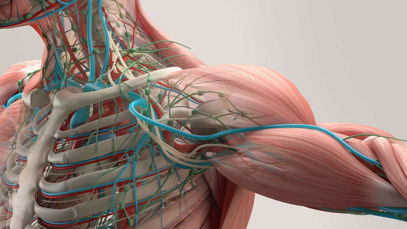
Prof. Dr. Murat Demirel
Orthopedics and Traumatology Specialist
Orthopedics Specialist Prof. Dr. Murat Demirel was born in Ankara in 1974. He completed his primary education at Ankara Kavaklıdere Primary School and his secondary and high school education at Ankara Atatürk Anatolian High School. Dr. Demirel graduated from Ankara University Faculty of Medicine in 1998 and completed his residency in Orthopedics and Traumatology at Ankara Numune Training and Research Hospital, 1st Orthopedics and Traumatology Clinic, in 2004.
PhD
Ankara University Institute of Health Sciences
Specialization
Ankara Numune Training and Research Hospital, 1st Orthopedics Clinic
Medical School
Ankara University Faculty of Medicine
Yazı İçeriği
What Does Calcific Tendinitis Mean and Where Is It Most Commonly Seen?
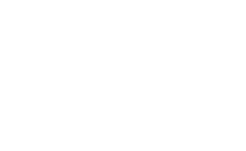
To better understand calcific tendinitis, let’s briefly look at how the shoulder joint works. Our shoulder is the most mobile joint in the body. This complex structure, which allows us to move our arm in nearly every direction, is controlled by four important tendons that come together to form what we call the “rotator cuff.” These tendons act like a conductor, precisely coordinating arm movements. Calcific tendinitis occurs when calcium builds up inside these important tendons.
Although it can be seen in different tendons in the body (such as the hip or elbow), the vast majority of calcific tendinitis cases occur in the shoulder. In the shoulder, the most commonly affected area is the “supraspinatus” tendon, which is responsible for lifting the arm sideways and upwards. This condition usually affects individuals between the ages of 30 and 60 and is slightly more common in women than in men.
Here is another interesting detail: calcific deposits can also be found in the shoulder X-rays of completely healthy individuals with no shoulder complaints, taken for another reason. This shows us that the mere presence of calcium deposits does not always indicate a problem; additional triggering factors must come into play for pain and other symptoms to appear. In other words, these deposits can remain silently in your shoulder for years without causing any discomfort.
What Are the Causes Behind the Development of Calcific Tendinitis?
The question “Why did these calcium deposits form in my shoulder?” is one of the most common and valid concerns patients have. Although the exact cause of calcific tendinitis has not yet been clarified, there are several strong theories believed to trigger this process. Contrary to popular belief, this buildup is not directly related to a calcium-rich diet or dairy consumption. The problem is not how the body uses calcium, but rather a series of cellular events occurring in the tendon tissue.
The most widely accepted theory is that the cells in the tendon tissue undergo a kind of transformation (metaplasia). Some conditions thought to trigger this transformation include:
- Reduced blood flow to the tendon over time
- Decreased oxygen levels within the tendon (hypoxia)
- Repeated microtrauma and overuse
- Natural wear and tear of the tendon with aging
These factors disrupt the tendon’s normal healing mechanism and trigger an abnormal process in which calcium crystals accumulate in the area as part of the repair process.
It has also been observed that some people are more prone to calcific tendinitis. We can list the risk factors as follows:
- Diabetes
- Hypothyroidism (underactive thyroid gland)
- Metabolic and endocrine diseases such as hyperparathyroidism
- Genetic predisposition (family history of this condition)
The presence of these risk factors may predispose to calcification by affecting the body’s general inflammatory response and tissue healing capacity. Therefore, when planning treatment, the patient’s overall health condition and other chronic illnesses must always be taken into consideration.
What Are the Symptoms That Suggest the Presence of Calcific Tendinitis?
The symptoms of calcific tendinitis can vary greatly depending on the individual and the stage of the disease. While some patients manage with mild discomfort, for others it can cause pain so severe that it disrupts daily life.
The most common symptoms are:
- Sudden or gradually increasing severe shoulder pain
- Pain that worsens at night and interrupts sleep
- Inability to lie on the affected shoulder
- Difficulty raising the arm overhead or moving it backward
- Stiffness and limited mobility in the shoulder joint
- Difficulty performing daily tasks (dressing, combing hair, reaching for objects)
- Tenderness when touching the front or outer part of the shoulder
- Long-term weakness in the shoulder muscles
The nature of the pain also changes depending on the stage of the disease. In the “formation” phase, when calcium gradually accumulates, the pain is usually chronic, aching, and worsens with activity. However, in the “resorptive” phase, when the body perceives the deposit as a foreign substance and tries to remove it, a very severe inflammatory reaction occurs. The pain during this phase is often described as throbbing, stabbing, and unbearable. The onset of this severe pain is actually part of the body’s healing mechanism and may indicate that the deposit is beginning to dissolve, rather than being a sign of worsening.
Contact us for detailed information and an appointment!
Which Methods Are Used to Diagnose Calcific Tendinitis?
When you visit a doctor with shoulder pain, the first step toward correct treatment is to clearly identify the source of the pain. The diagnosis of calcific tendinitis is usually made through a multi-step approach.
Medical History and Physical Examination:
First, we listen to your complaints in detail. Questions such as when your pain started, its nature, which movements worsen it, and how it affects your sleep form the foundation of the diagnostic process. Then, a comprehensive shoulder examination is performed. In this exam, your shoulder’s range of motion, muscle strength, tender points, and special tests to rule out other possible conditions like shoulder impingement are assessed.
Imaging Methods:
After the physical examination, we use imaging methods to confirm our suspicions and clarify the condition.
The information provided by imaging methods includes:
- Definitively confirming the presence of calcium deposits
- Identifying which tendon contains the deposit
- Determining the size, shape, and number of deposits
- Understanding the structure of the deposit (hard or soft)
- Determining the stage of the disease (formation or resorption)
- Evaluating whether there are other accompanying problems (tendon tears, bursitis, etc.)
The main imaging methods we use for this purpose are:
X-ray: The first and most basic method in diagnosing calcific tendinitis. It shows calcium deposits very clearly and is usually sufficient for diagnosis.
Ultrasound (USG): A radiation-free, dynamic, and highly sensitive method. Ultrasound provides more detailed information about the structure of the deposits compared to X-ray. It allows us to distinguish between “toothpaste-like” soft deposits and “rock-like” hard ones. This distinction is particularly important when planning treatments such as needle lavage.
Magnetic Resonance Imaging (MRI): Not routinely required for diagnosing calcific tendinitis. However, when the diagnosis is uncertain or if there is suspicion of complex conditions such as rotator cuff tears, MRI may be requested to evaluate the surrounding soft tissues and joint in more detail.
It is also important to rule out other conditions that may cause shoulder pain. Symptoms of calcific tendinitis can sometimes overlap with other disorders.
Some conditions considered in differential diagnosis include:
- Shoulder impingement syndrome
- Frozen shoulder (adhesive capsulitis)
- Rotator cuff tendon tears
- Acromioclavicular joint arthritis
- Referred pain from cervical disc herniation
The correct diagnosis is made after evaluating all these possibilities, ensuring the creation of the most effective personalized treatment plan.
What Are the First-Line Treatment Approaches for Calcific Tendinitis?
In treating calcific tendinitis, the first step is almost always non-surgical, that is, conservative methods. Most patients achieve significant relief with these approaches. The main goal is to control pain, reduce inflammation, and preserve shoulder function.
The initial treatment methods include:
Activity Modification and Rest: Avoiding movements that trigger pain, especially overhead and strenuous activities, allows the inflamed tendon to rest and recover. This does not mean keeping the shoulder completely immobile, but rather using it within the limits of pain.
Cold and Heat Applications: In the acute phase, when pain is very severe and new, applying cold packs to the affected area several times a day for 15–20 minutes can help reduce pain and inflammation. In more chronic cases or to relax the muscles, heat application may also be beneficial.
Medication: Non-steroidal anti-inflammatory drugs (NSAIDs), used under medical supervision, help control symptoms with both pain-relieving and anti-inflammatory effects.
Physical Therapy and Rehabilitation: One of the cornerstones of treatment that ensures long-term success. A program tailored to the patient and conducted under the supervision of a physiotherapist serves multiple purposes.
The main goals of physical therapy are:
- Reducing pain and inflammation
- Maintaining and improving shoulder range of motion
- Preventing “frozen shoulder”
- Strengthening the muscles around the shoulder and scapula
- Teaching correct posture and shoulder usage habits
As part of the physical therapy program, treatment begins with passive stretching exercises and gradually progresses to active movements and strengthening exercises. Commonly used exercises include pendulum exercises, wall climbing, and resistance band training. This process requires patience but is critical for restoring shoulder health.
What Is the Role of Injection Methods in the Treatment of Calcific Tendinitis?
When initial treatments fail to control pain, especially severe pain that disrupts sleep, injection therapies may be considered.
Corticosteroid (Cortisone) Injections: Cortisone is a very powerful anti-inflammatory drug. When injected into the bursa (subacromial bursa) above the calcium deposit or directly into the joint, it can rapidly suppress the intense inflammatory response in the area, providing significant pain relief. These injections can open a “window” for rehabilitation, especially in patients unable to perform physical therapy due to severe pain. However, cortisone’s effect is usually temporary and does not eliminate the underlying calcium deposit. Therefore, it is generally considered part of the treatment rather than a permanent solution. Repeated injections must be limited and performed with caution due to potential tendon damage.
Ultrasound-Guided Lavage and Barbotage (Needle Washout): This method serves a different purpose than cortisone injections; it not only suppresses symptoms but directly targets the source of the problem. Under ultrasound guidance, a needle is inserted into the calcium deposit. The deposit is first mechanically broken up with the needle (barbotage), then sterile fluid is injected and withdrawn to flush out chalky calcium particles (lavage). This procedure is performed under local anesthesia in an outpatient setting and is a minimally invasive alternative to surgery. It is particularly effective in soft, “toothpaste-like” deposits and can stimulate the body’s natural healing cells to accelerate absorption of the remaining deposit.
Is Extracorporeal Shock Wave Therapy (ESWT) an Effective Method for Calcific Tendinitis?
In persistent calcific tendinitis cases unresponsive to other conservative methods, another effective and modern treatment before resorting to surgery is Extracorporeal Shock Wave Therapy (ESWT). Similar to the technology used in breaking kidney stones, this treatment works by focusing high-energy sound waves from outside the body onto the calcium deposit.
ESWT’s mechanism of action has two aspects.
Mechanical Effect: The shock created by the sound waves breaks hard calcium deposits into smaller pieces. These smaller fragments are more easily absorbed by the body’s natural cleaning mechanisms.
Biological Effect: The treatment is not just a mechanical process. It also triggers a biological response in the tendon tissue. It increases blood flow (neovascularization), stimulates the release of growth factors that promote tissue healing and regeneration, and reduces chronic inflammation, thereby providing pain relief.
ESWT treatment usually consists of several sessions performed weekly. Although mild discomfort may be felt during the procedure, anesthesia is not required. Particularly in harder and more chronic deposits, ESWT produces highly successful results and can save many patients from surgical intervention.
When Is Surgery Considered for Calcific Tendinitis?
Surgery is always the last option in the treatment of calcific tendinitis. The vast majority of patients recover with non-surgical methods. However, in some cases, surgery becomes the most appropriate and effective solution.
The situations that require surgical treatment include:
- Persistent pain and symptoms despite all non-surgical treatments (at least 6 months)
- Pain severely affecting the patient’s quality of life (sleep, daily activities, work)
- Progressive restriction of shoulder movement and loss of strength
- Very large calcium deposits causing mechanical impingement or catching in the shoulder
Today, calcific tendinitis surgery is performed with the arthroscopic (minimally invasive) method, considered the “gold standard.” Using 2–3 very small (about 1 cm) incisions, a camera and special surgical instruments are inserted into the shoulder without opening the joint. Under magnified visualization, the calcium deposit is located, carefully cleaned, and the area thoroughly irrigated. If a significant tendon tear occurs where the calcium was removed, it can also be repaired arthroscopically during the same procedure.
The advantages of arthroscopic surgery are numerous:
- Less postoperative pain
- Faster recovery process
- Lower risk of infection
- Better cosmetic results
Success rates after surgery are quite high. However, the success of the surgery depends on the patient’s full adherence to the postoperative physical therapy and rehabilitation program, which usually lasts several months.
What Happens If Calcific Tendinitis Is Left Untreated or Neglected?
Although calcific tendinitis is usually a benign condition that can resolve spontaneously over time, this process can take years, and the associated pain and functional loss may become permanent. Untreated or neglected calcific tendinitis can lead to some undesirable complications.
Possible complications include:
- Chronic shoulder pain
- Development of frozen shoulder (adhesive capsulitis)
- Permanent limitation of movement
- Persistent weakness and atrophy of the rotator cuff muscles
- In rare cases, tendon rupture due to weakening caused by large deposits
The best way to prevent these complications is to consult a specialist as soon as shoulder pain begins and manage the condition appropriately.
What Can Be Done to Prevent Recurrence of Calcific Tendinitis?
Although there is no magical formula to completely prevent the development of calcific tendinitis, there are certain measures that can be taken to protect shoulder health and reduce the risk.
Preventive approaches and recommendations include:
- Avoiding repetitive and overhead activities that strain the shoulder (such as professional swimming, volleyball, or painting) or taking regular breaks during these activities
- Not lifting heavy loads suddenly or uncontrollably
- Gradually and progressively increasing intensity when starting a new sport or exercise program
- Performing regular exercises that strengthen and increase the flexibility of the shoulder and back muscles
- Making ergonomic adjustments in office settings (correct desk and chair height, monitor position, etc.)
- Ensuring regular control and treatment of underlying chronic conditions such as diabetes
Remember that the best method of prevention is to listen to your body’s signals and not ignore warning signs such as pain. With the right approaches, calcific tendinitis is generally a condition that does not leave permanent problems in the long term, and patients can return to their normal lives with great satisfaction.
Contact us for detailed information and an appointment!
Blog Posts
What Is a Tendon Rupture? Symptoms, Causes, and Treatment Methods
Ankara Ortopedi | Prof. Dr. Murat Demirel » General » What Is a Tendon Rupture? [...]
13
Apr
Apr
What Is an Orthosis? Why Is an Orthosis Used?
Ankara Ortopedi | Prof. Dr. Murat Demirel » General » What Is an Orthosis? Why [...]
13
Apr
Apr

Knee Replacement Surgery Prices
Ankara Ortopedi | Prof. Dr. Murat Demirel » Knee Treatments » Knee Replacement Surgery PricesThe [...]
18
Aug
Aug
Hip Replacement Surgery Prices
Ankara Ortopedi | Prof. Dr. Murat Demirel » Hip Treatments » Hip Replacement Surgery PricesThe [...]
18
Aug
Aug