


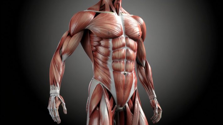
Prof. Dr. Murat Demirel, one of the best orthopedic doctors in Ankara specializing in hip impingement syndrome treatment, stands out with his many years of experience in the diagnosis and treatment of this condition, which occurs as a result of bone structures in the hip joint rubbing against each other. Hip impingement syndrome is especially common in young and active individuals, presenting with pain, limited movement, and stiffness in the joint during sports or daily activities. If left untreated, it may lead to cartilage damage and early osteoarthritis. Prof. Dr. Demirel offers personalized treatment plans in advanced medical facilities with high hygiene standards, among the hospitals in Ankara that provide hip impingement syndrome treatment.
Depending on the severity of the disease, conservative methods such as rest, physical therapy, PRP, or stem cell injections can be applied during the treatment process. In advanced cases, hip arthroscopy can be used to correct bone protrusions. Following hip impingement syndrome treatment, the goal is to achieve a fast, safe, and lasting recovery with the doctor’s recommendations. In addition, transparent and up-to-date information about hip impingement syndrome treatment costs in Ankara is provided to help patients make informed decisions. You can also protect your hip health, reduce pain, and increase your freedom of movement by contacting us immediately to book an appointment.
| Disease Name | Hip Impingement (Femoroacetabular Impingement – FAI) |
| Affected Area | Hip joint (between femoral head and acetabulum) |
| Types | Cam type, Pincer type, Combined type |
| Symptoms | Groin and hip pain, limited movement, clicking sound, pain when climbing stairs |
| Diagnostic Methods | Physical examination, X-ray, MR arthrography, CT scan |
| Causes | Anatomical abnormalities, sports activities, hip developmental disorders |
| Risk Factors | Intensive sports, genetic predisposition, hip trauma |
| Treatment Methods | Physical therapy, drug therapy (NSAIDs), intra-articular injection, arthroscopic surgery |
| Surgical Option | Correction of bone irregularities with hip arthroscopy |
| Complications | Hip stiffness, cartilage damage, development of arthritis, nerve damage |
| Recovery Process | Weeks with conservative treatment, 3–6 months after surgery |
| Prevention Methods | Avoiding excessive strenuous sports, proper warm-up, early diagnosis |


Prof. Dr. Murat Demirel
Orthopedics and Traumatology Specialist
Orthopedics Specialist Prof. Dr. Murat Demirel was born in Ankara in 1974. He completed his primary education at Ankara Kavaklıdere Primary School and his secondary and high school education at Ankara Atatürk Anatolian High School. Dr. Demirel graduated from Ankara University Faculty of Medicine in 1998 and completed his residency in Orthopedics and Traumatology at Ankara Numune Training and Research Hospital, 1st Orthopedics and Traumatology Clinic, in 2004.
PhD
Ankara University Institute of Health Sciences
Specialization
Ankara Numune Training and Research Hospital, 1st Orthopedics Clinic
Medical School
Ankara University Faculty of Medicine
Yazı İçeriği
What is Hip Impingement Syndrome?
To understand this syndrome, let’s first take a look at the magnificent design of the hip joint. The hip joint is one of the strongest and most mobile joints in the body. We can compare its structure to a ball and socket; just like a ball rotating inside a bowl. The “ball” part is the spherical head at the upper end of our thigh bone (femur). The “socket” is the hollow part of our pelvis where this ball fits, medically known as the acetabulum.
The surfaces where these two bones come into contact are covered with joint cartilage, a smooth, shiny, and slippery tissue. Despite being millimeters thin, this cartilage performs an incredible task; it allows the bones to move without friction, pain, or stiffness. In addition, there is a very special structure called the “labrum” that surrounds the edge of the socket. The labrum is a strong and flexible ring of cartilage. We can compare it to the rubber seal on the lid of a jar. Its functions are critical: it deepens the socket to keep the ball more stable, balances the pressure inside the joint, and prevents leakage of joint fluid, helping nourish the cartilage.
So how does Hip Impingement Syndrome disrupt this perfect mechanism? The problem lies in the shape of the bones. Normally, the ball and socket should work in perfect harmony, but due to congenital differences or structural variations that develop during adolescence, this harmony is disrupted. Either the ball is not perfectly spherical and has a bump, or the socket covers the ball more than it should, acting like a clamp.
These extra bone formations collide with and pinch other joint structures during certain movements (especially flexion and rotation). These repetitive microtraumas eventually cause tears in the precious labrum and irreversible wear of the cartilage. In short, this syndrome is essentially a problem of mechanical friction and impact.
How Many Types of Hip Impingement Syndrome Are There?
Depending on the source of the impingement, that is, where the bony problem is located, we classify this syndrome into three main types. Making this distinction is vital to drawing the most accurate, personalized treatment plan.
Cam Type Impingement
Here, the problem lies in the “ball,” i.e., the femoral head. Instead of being a smooth sphere, the femoral head has an extra bony bump where the head and neck meet. As the hip moves, this bump strikes the rim of the socket. This is like a marble with dried glue on it trying to roll smoothly; it will snag and scratch the surface with every turn. Similarly, this Cam-type bump scrapes and wears down the labrum and cartilage with every movement. It is more commonly seen in young and active men.
Pincer Type Impingement
In this type, the problem lies in the “socket,” i.e., the acetabulum. The rim of the socket is deeper than normal or has a bony protrusion. This causes the socket to cover the ball excessively, acting like a clamp. When the hip reaches the end of its motion, the femoral neck strikes this excessive coverage, compressing the labrum in between. We can compare this to a teacup rim being too deep and crushing the teabag every time you stir the spoon. This type mainly causes labrum damage and is more common in middle-aged women.
Combined (Mixed) Type Impingement
In fact, this is the most common situation in clinical practice. Most patients have both Cam type (bump on the ball) and Pincer type (extra coverage in the socket) deformities together. This means that the joint is attacked by two mechanisms at once, making the damage more complex and requiring correction of both problems during surgical treatment.
Causes and Risk Factors of Hip Impingement Syndrome
One of the most common questions patients ask is, “Why did this happen to me?” The fundamental cause of Hip Impingement Syndrome is that the hip bones do not develop into their ideal shape during adolescence due to a combination of genetic and mechanical factors. This is largely beyond a person’s control. In other words, it’s not your fault.
However, not everyone with this bone structure develops symptoms. Certain factors increase the risk of this silent condition turning into a painful syndrome. These risk factors include:
- Intense sports activities
- History of childhood hip disorders
- Genetic predisposition
- Occupational strain
- Previous hip trauma
Let’s expand on these factors. Sports that involve continuous and extreme hip flexion and rotation are the most important triggers. Athletes engaged in football, ice hockey, basketball, ballet, martial arts, and weightlifting who also have underlying bone abnormalities are more likely to develop symptoms earlier. It is important to remember that sports do not cause this syndrome; they simply reveal an existing structural problem. Similarly, some childhood hip diseases (such as Legg-Calve-Perthes or slipped capital femoral epiphysis) can alter the shape of the femoral head, paving the way for Cam-type impingement later in life.
Contact us for detailed information and an appointment!
Which Symptoms Should Suggest Hip Impingement Syndrome?
The symptoms of Hip Impingement Syndrome usually develop gradually and can significantly reduce a person’s quality of life over time. If one or more of the following symptoms sound familiar to you, it is advisable to consult a specialist.
The most common symptoms are:
- Groin pain
- Pain on the side of the hip
- A deep, dull aching sensation
- Sharp and stabbing pain
- Sensation of catching or locking
- A “click” sound from the hip
- Limited movement
- Limping
- Increased discomfort while sitting
- Decreased sports performance
The most typical symptom is groin pain. Patients often describe the pain by cupping their hand in the shape of a “C” around the front and side of the hip; we call this the “C sign,” which is quite characteristic for impingement syndrome. The pain often appears after prolonged sitting (for example, after a long car journey or an office meeting), when getting up from a low chair, bending down to put on socks, or getting in and out of a car.
During activity, the character of the pain may change; sudden twisting or squatting movements may cause sharp, stabbing pain. Mechanical symptoms such as catching, locking, or a “click” sound are also common. These mechanical symptoms often result from a torn labrum or a loose piece of cartilage getting trapped in the joint. Over time, the body restricts movement to avoid pain, leading to stiffness and limited mobility in the hip joint, particularly in inward rotation.
Which Methods Are Used to Diagnose Hip Impingement Syndrome?
Accurate diagnosis is the first and most important step in effective treatment. We do not rely on a single finding or a single scan to diagnose Hip Impingement Syndrome. Like a detective, we piece together all the clues to reach the final conclusion. This process consists of several essential steps.
The diagnostic methods we use include:
- Detailed medical history
- Comprehensive physical examination
- Special X-ray imaging
- Magnetic Resonance Imaging (MRI / MR Arthrography)
- Computed Tomography (CT)
- Diagnostic intra-articular injection
First, we listen to you carefully. Information such as when, where, and how your pain started, what movements make it better or worse, your lifestyle, and the sports you engage in are very valuable for us. Then, we move on to a physical examination. We assess your gait, posture, hip range of motion, and muscle strength. The key part of this examination is impingement tests. The most commonly used is the FADIR test, where, lying on your back, we bend your hip and knee and then rotate your leg inward. If this movement triggers that familiar groin pain, suspicion of impingement syndrome increases significantly.
Imaging methods confirm the diagnosis. The first step is always X-rays taken in special positions. X-rays perfectly show the bone structure and allow us to identify Cam or Pincer type abnormalities. With special measurements on these images (such as alpha angle, lateral center-edge angle), we can determine the type and degree of impingement.
After evaluating the bones, we move on to the soft tissues, namely the labrum and cartilage. The best method for assessing these is MRI. Particularly, MR Arthrography with contrast injection into the joint clearly shows even the smallest tears in the labrum and the extent of cartilage damage. In some complex cases, or for surgical planning, we may also use CT scans to view the bone structure in 3D.
Finally, sometimes we use diagnostic injections to ensure the pain is not referred from the back or another source. If local anesthetic injected directly into the hip joint provides significant relief, we confirm that the hip joint is indeed the source of the problem.
Can Hip Impingement Syndrome Be Treated Without Surgery?
Yes, not every case of Hip Impingement Syndrome requires immediate surgery. Especially if there is no significant cartilage damage and the symptoms can be controlled, our first choice is always non-surgical (conservative) treatments. The goal of these treatments is to relieve pain, improve function, and protect the joint from further damage.
Non-surgical treatment options include:
- Activity modification
- Medication
- Physical therapy and rehabilitation
- Intra-articular injections
The first step in treatment is to identify and avoid or adjust activities that trigger pain. For example, raising the bike saddle, switching to a higher office chair, or avoiding deep squatting exercises can make a big difference. To relieve pain and inflammation, nonsteroidal anti-inflammatory drugs (NSAIDs) may be prescribed under medical supervision. However, the cornerstone of treatment is physical therapy. A well-planned physical therapy program not only relieves symptoms but also strengthens the hip muscles and increases flexibility, improving joint mechanics.
If pain persists despite these methods, a cortisone injection into the joint may be considered. Cortisone is a powerful anti-inflammatory that can reduce joint inflammation and provide relief lasting several months. While this does not solve the underlying bone problem, it serves as a “bridge,” helping the patient get through the painful period and participate more effectively in physical therapy.
When and How is Surgical Treatment Necessary for Hip Impingement Syndrome?
If, despite at least 3–6 months of regular non-surgical treatment (especially a well-structured physical therapy program), your pain persists, your quality of life decreases, and imaging reveals structural damage such as a labrum tear, surgical treatment becomes the best option. The decision for surgery is made jointly with the patient, considering factors such as age, activity expectations, and the degree of joint damage.
The main goals of surgery are:
- Trimming excessive bone causing impingement
- Repairing torn labrum
- Repairing damaged cartilage
- Restoring normal, pain-free joint movement
- Slowing down the development of arthritis in the long term
Today, this surgery is most commonly performed using a minimally invasive method called hip arthroscopy. Instead of making a large incision, several small incisions of about 1 cm are made. A camera is inserted through one incision, projecting the inside of the joint onto a large screen, while miniature surgical instruments are inserted through the others. The surgeon uses these instruments to shave down the bony bump (Cam lesion) or the socket overcoverage (Pincer lesion). Torn labrum tissue is reattached to the rim of the socket with small anchors and special sutures. Whenever possible, repairing the labrum is the best option for the joint’s long-term health.
The advantage of this minimally invasive technique is that it causes much less tissue damage compared to open surgery, reduces postoperative pain, shortens hospital stay, and allows a much faster return to daily life.
How Does Recovery Progress After Hip Impingement Syndrome Surgery?
A successful surgery is only half of the equation. The other half is careful and patient rehabilitation after the operation. This process allows repaired tissues to heal while preventing stiffness and muscle weakness. Although recovery speed varies from patient to patient, the rehabilitation protocol generally consists of certain stages.
Phase 1: Early Protection Phase (0–4 Weeks After Surgery)
The sole purpose of this period is protection. The repaired labrum and other tissues are very delicate.
Things to Do:
- Use crutches (usually for the first 2–4 weeks)
- Apply controlled and limited weight on the operated leg
- Start gentle, passive movements with the help of a physiotherapist
- Apply ice therapy
- Perform simple isometric exercises as instructed
Avoid:
- Sudden and forceful movements
- Crossing legs
- Deep squats or sitting on low chairs
- Actively lifting the leg straight
Phase 2: Intermediate Phase (4–12 Weeks After Surgery)
Now it’s time to gradually regain movement and strength.
Things to Do:
- Gradually stop using crutches
- Work on regaining full range of motion
- Begin strengthening hip muscles (bridge, clamshell, etc.)
- Start balance exercises
- Engage in low-impact cardio such as stationary cycling
Avoid:
- Running and jumping
- Pushing through exercises that cause pain
- Lifting heavy weights
Phase 3: Advanced Phase and Return to Sport (3–6 Months and Beyond)
The final stage aims to safely return the patient to their previous activity level.
Things to Do:
- Progress to more challenging strengthening exercises
- Gradually begin a running program
- Start agility and sport-specific drills
Avoid:
- Returning to full-speed sports without sufficient strength and control
- Skipping warm-up and cool-down routines
Full return to sports usually takes 6 months, sometimes longer. Patience and listening to the body’s signals are the most important rules in this process.
Can Hip Impingement Syndrome Treatment Prevent Osteoarthritis?
This is perhaps the most important long-term goal of treatment. If left untreated, Hip Impingement Syndrome causes ongoing abnormal friction in the joint, gradually destroying the cartilage over the years, ultimately resulting in early hip osteoarthritis. Once hip arthritis progresses, the only solution may be hip replacement surgery.
With timely surgical intervention to eliminate the bone problem and repair the torn labrum, this destructive process inside the joint can be halted. This slows or prevents cartilage damage from progressing. Scientific studies have shown that hip arthroscopy performed before cartilage damage advances is highly successful in preserving the joint and delaying or completely preventing the need for hip replacement. Therefore, this treatment not only relieves today’s pain but also serves as an important investment in the future of your hip.
Contact us for detailed information and an appointment!
Frequently Asked Questions
What is hip impingement syndrome and how does it occur?
Hip impingement syndrome (femoroacetabular impingement – FAI) is a condition where abnormal contact between the bones of the hip joint leads to damage of the joint cartilage and labrum. It usually develops due to congenital hip shape abnormalities, repetitive sports activities, or trauma.
What are the symptoms?
Pain in the groin or front of the thigh, stiffness in the hip, limited range of motion, and increased discomfort especially while sitting or bending are the main symptoms. Pain may worsen after sports or long walks. Sometimes catching or clicking sounds may be heard from the joint.
Who is more likely to develop it?
It is more common in young and active individuals, athletes (such as those engaged in football, basketball, or ballet involving intense hip movement), and in people with congenital hip structural abnormalities.
How is it diagnosed?
Physical examination reveals limited and painful hip movements. To confirm the diagnosis, X-rays, magnetic resonance imaging (MRI), or computed tomography (CT) are used. MRI provides detailed evaluation of the labrum and cartilage.
What treatment methods are used for hip impingement syndrome?
In the early stages, rest, activity modification, pain relief medication, physical therapy, and exercise programs are applied. In resistant or advanced cases, arthroscopic surgery can be performed to correct bone abnormalities and repair the labrum.
When is surgery necessary?
Surgical treatment is recommended for patients with severe pain and significant restriction of hip movements that do not improve with physical therapy and lifestyle changes and that affect daily life.
What is the recovery process like after surgery?
In the first days after surgery, crutches are used. With physical therapy, muscle strength and range of motion improve. Full recovery and return to sports usually take between 2–4 months. Recovery varies depending on the procedure performed and patient compliance.
What happens if hip impingement syndrome progresses?
If left untreated, hip impingement syndrome may lead to joint arthritis, chronic pain, and permanent loss of movement over time. Therefore, early diagnosis and treatment are important.
Why are exercise and physical therapy important in hip impingement syndrome?
Exercises that strengthen hip muscles and increase flexibility help reduce pain, preserve joint mobility, and decrease the need for surgery. Programs supervised by a physiotherapist provide better results.
Can hip impingement syndrome be prevented?
Although genetic and structural factors cannot be prevented, practicing appropriate exercises at a young age, avoiding excessive strain and repetitive trauma, maintaining strong muscles, and taking care of hip health can help reduce the risk.
Blog Posts
What Is a Tendon Rupture? Symptoms, Causes, and Treatment Methods
Ankara Ortopedi | Prof. Dr. Murat Demirel » General » What Is a Tendon Rupture? [...]
13
Apr
Apr
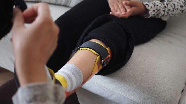
What Is an Orthosis? Why Is an Orthosis Used?
Ankara Ortopedi | Prof. Dr. Murat Demirel » General » What Is an Orthosis? Why [...]
13
Apr
Apr

Knee Replacement Surgery Prices
Ankara Ortopedi | Prof. Dr. Murat Demirel » Knee Treatments » Knee Replacement Surgery PricesThe [...]
18
Aug
Aug
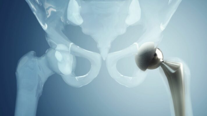
Hip Replacement Surgery Prices
Ankara Ortopedi | Prof. Dr. Murat Demirel » Hip Treatments » Hip Replacement Surgery PricesThe [...]
18
Aug
Aug