

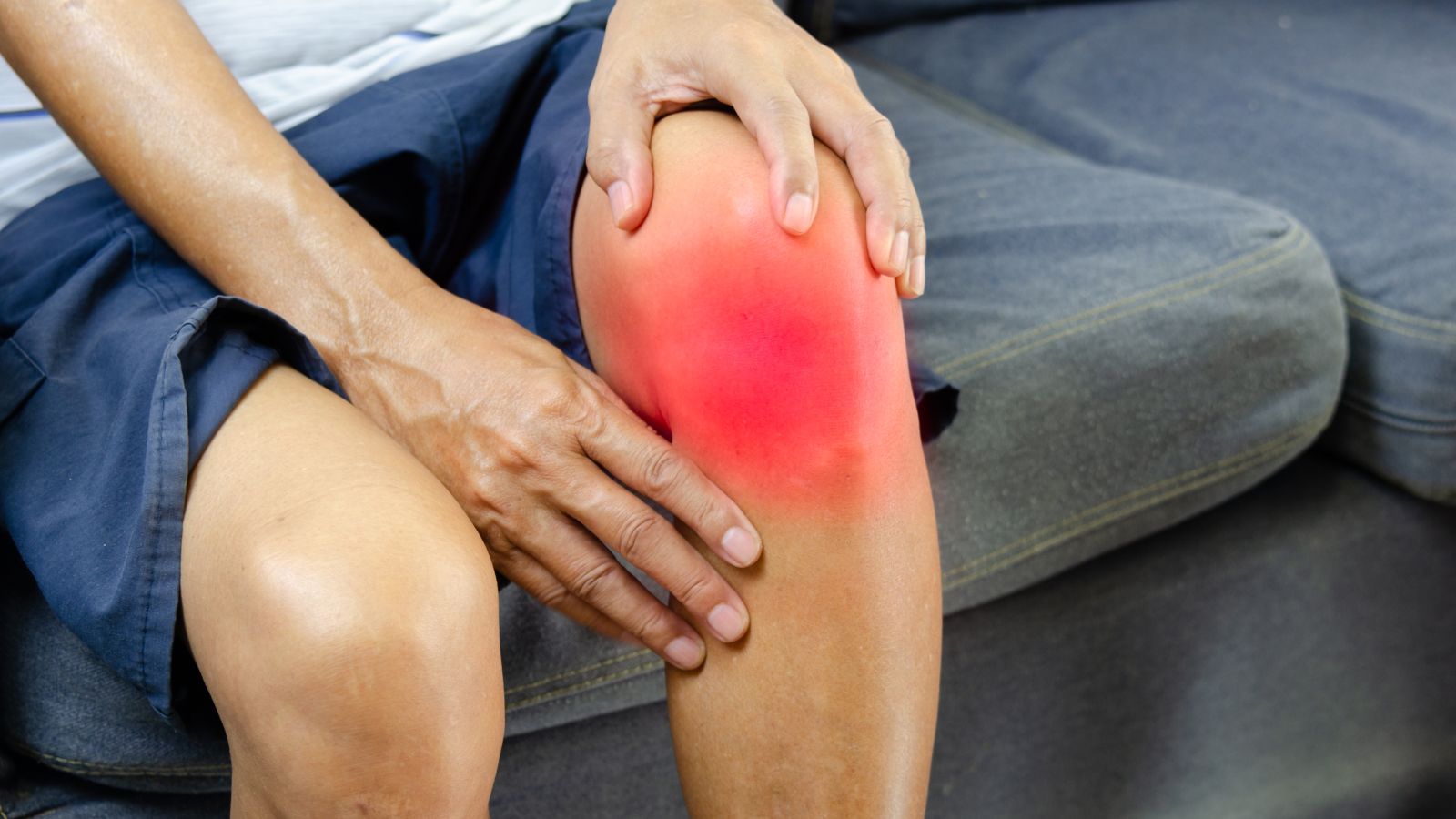
Prof. Dr. Murat Demirel, one of the best orthopedic surgeons performing patellar dislocation surgery in Ankara, stands out with his long years of experience in treating this problem that disrupts the stability of the knee joint and can lead to recurrent dislocations. Patellar (kneecap) dislocation usually occurs due to trauma, sudden twisting movements, or laxity in the ligamentous structures, and if not treated, it may result in cartilage damage and permanent problems in the joint. Among the hospitals in Ankara that perform patellar dislocation surgery, Prof. Dr. Demirel offers personalized surgical plans for his patients in centers with advanced medical equipment and high hygiene standards.
Before surgery, Prof. Dr. Demirel examines the knee joint structure in detail with advanced imaging methods and determines the most appropriate surgical technique to restore joint stability. By following patients closely after patellar dislocation surgery and providing clear medical recommendations, he helps the recovery process proceed quickly, safely, and permanently. In addition, transparent and up-to-date information is provided on patellar dislocation surgery costs in Ankara to help patients make informed decisions. To protect your knee health, reduce your pain, and increase your freedom of movement, you can contact us now to book an appointment.
| Disease Name | Patellar Dislocation |
| Affected Area | Articular surface between the patella (kneecap) and the femur |
| Symptoms | Sudden knee pain, lateral displacement of the patella, swelling, restricted movement, sensation of giving way |
| Diagnostic Methods | Clinical examination, X-ray, MRI (to assess ligament tears and cartilage damage) |
| Causes | Trauma, sudden twisting movements, congenital patellar instability |
| Risk Factors | Young age, female sex, structural abnormalities in the knee, generalized ligamentous laxity |
| Treatment Methods | Conservative (immobilization, physical therapy), surgical (MPFL reconstruction, lateral release) |
| Surgical Options | Medial patellofemoral ligament (MPFL) reconstruction, trochleoplasty |
| Complications | Recurrent dislocation, cartilage damage, knee instability, arthritis |
| Recovery Process | 6–8 weeks after conservative treatment; 3–6 months after surgery |
| Prevention Methods | Balance and strength exercises, strengthening of the quadriceps muscles |
| Follow-up | Regular clinical controls and continuation of rehabilitation programs |


Prof. Dr. Murat Demirel
Orthopedics and Traumatology Specialist
Orthopedics Specialist Prof. Dr. Murat Demirel was born in Ankara in 1974. He completed his primary education at Ankara Kavaklıdere Primary School and his secondary and high school education at Ankara Atatürk Anatolian High School. Dr. Demirel graduated from Ankara University Faculty of Medicine in 1998 and completed his residency in Orthopedics and Traumatology at Ankara Numune Training and Research Hospital, 1st Orthopedics and Traumatology Clinic, in 2004.
PhD
Ankara University Institute of Health Sciences
Specialization
Ankara Numune Training and Research Hospital, 1st Orthopedics Clinic
Medical School
Ankara University Faculty of Medicine
Yazı İçeriği
What is a patellar dislocation and how is the knee’s delicate balance disrupted?
To grasp the answer, we need to appreciate what an engineering marvel the knee is. Our kneecap—patella—is the largest sesamoid bone in the body, embedded within the thick tendon of the powerful four-headed muscle at the front of the thigh (quadriceps). Its primary function is to act like a lever, boosting the force of this massive muscle when we straighten the leg and making movement more efficient.
Now imagine the very bottom end of the femur (thigh bone). There you’ll find a special V-shaped groove—like a set of train tracks—designed for the patella to glide safely as we bend and straighten the knee. We call this the “trochlear groove.” In a healthy knee, the patella moves up and down smoothly within this track. The outer ridge of this groove is typically higher than the inner ridge, serving as the first and most important bony barrier that prevents the kneecap from slipping outward.
A patellar dislocation is the event in which the kneecap bone, most commonly outward, completely leaves this track and goes off course.
But it isn’t only this bony track that keeps the patella in line. The knee’s delicate balance relies on the coordinated teamwork of several key stabilizers. If any member of this team weakens or fails to do its job, balance is lost and dislocation becomes likely. The most important of these are:
Medial Patellofemoral Ligament (MPFL): This very strong ligament extends from the inner edge of the patella to the femur. Think of it as the primary “seat belt” preventing the patella from sliding outward. It is the most critical structure for stability in the early degrees of bending (0–30°). In almost every dislocation, this ligament is either excessively stretched or completely torn.
Dynamic Muscle Support: The quadriceps in front of the thigh—and especially the segment known as the Vastus Medialis Obliquus (VMO), which practically works “for” the patella—are dynamic stabilizers that keep the kneecap aligned during movement. Balanced and timely contraction of these muscles prevents the patella from derailing.
Other Soft Tissues: The retinacula on each side of the patella also contribute to this stability network.
Why does a first patellar dislocation occur?
A first-time patellar dislocation is usually triggered by an acute event. Sometimes, however, because of an underlying structural issue the person may not even be aware of, it can occur with a much simpler, even non-traumatic, movement. The main causes fall into two groups:
Traumatic injuries: Typically due to a clear external force.
- A direct blow to the knee—for example, an opponent’s challenge in a football match or hitting the knee hard on an object.
- Falling directly onto the knee, creating a force that can push the patella out of its track.
Non-contact injuries: In fact, most dislocations occur without a direct blow, during the body’s own motion.
- The most common mechanism is when the foot is planted on the ground while the body suddenly rotates over the knee.
- A quick pivot in basketball.
- Twisting on the supporting leg while kicking in football.
- Sudden, uncontrolled moves in dance or gymnastics.
During such non-contact events, the knee is forced into a “knock-knee” (valgus) position and the patella slides outward. When this happens, not only does the bone move—almost always that critical seat belt, the MPFL, tears. In addition, as the patella dislocates and then relocates, the cartilage on the undersurface of the patella can collide with the edge of the femoral groove. This impact may cause small pieces of cartilage and bone to break off (osteochondral fractures). These fragments can float freely inside the joint, causing pain, catching, and, in the long run, significant joint damage.
Remember: trauma is often the last drop that overflows the cup. How easily a dislocation happens usually depends on a pre-existing “weak link” in the knee’s structure. A knee with robust, normal anatomy requires a substantial trauma to dislocate, whereas in those with underlying predispositions, much simpler movements can trigger a dislocation.
Why is patellar dislocation prone to recur?
After the first dislocation, the risk of recurrence increases dramatically. That’s because the initial event causes lasting damage to the stability mechanisms and certain anatomical risk factors may fuel the problem. The key reasons are:
- MPFL insufficiency: The number one cause of recurrent dislocations. Once torn or overstretched, the MPFL rarely heals at the correct length and tension. If it remains lax or dysfunctional, the knee loses its most important passive restraint. Like a car seat with a broken seat belt, the patella becomes prone to slipping out with even minor jolts.
- Trochlear dysplasia (shallow/flat groove): In some individuals, the V-shaped groove where the patella tracks is congenitally too shallow—or even flat. The shallower the groove, the less bony containment the patella has, making outward slippage much easier. This is one of the most significant anatomical risk factors.
- Patella alta (high-riding patella): The kneecap sits higher than normal. As the knee begins to bend, the patella engages the bony groove later than it should. In that early, most vulnerable range of motion, the patella lacks bony support and can more easily lose balance.
- Increased TT–TG distance (abnormal pull vector): A technical measurement indicating that the attachment point of the patellar tendon on the shin bone is more lateral than normal relative to the femoral groove. When this distance is increased, quadriceps contraction creates a vector pulling the patella outward, keeping it under constant lateral stress—crucial to consider in surgical planning.
- Malalignment of the limb: “Knock-knee” (genu valgum) alters the mechanical axis, increasing the load and outward pull on the patella.
- Generalized ligamentous laxity (hypermobility): Some people have inherently more elastic and lax ligaments throughout the body, which increases the tendency for joint instabilities, including dislocations in the shoulder or elbow.
- Muscle imbalances and weakness: Discoordination among stabilizing muscles is a key factor. A weak VMO (which pulls the patella medially) or, conversely, a tight lateral retinaculum that overpulls laterally both shift balance outward.
Several of these factors often coexist and add up. Each dislocation event further damages already weak structures and a vicious cycle begins: Dislocation → Ligament & cartilage injury → Increased laxity & instability → Lower force needed for recurrence → More structural damage. The main objective of treatment is to break this dangerous cycle and restore lasting stability.
Contact us for detailed information and an appointment!
What symptoms appear with a patellar dislocation and how is it recognized?
The symptoms are quite characteristic and are usually easily noticed by the patient. It’s best to consider them in two groups: the “acute” first-time event and the “recurrent/chronic” situation.
Symptoms during an acute patellar dislocation:
- Sudden, stabbing, severe pain
- A clear “pop” or snapping sensation heard/felt in the knee
- A visible deformity of the knee
- The patella appearing as a bulge on the outer side of the knee
- A distinct feeling that the kneecap has slipped out of place
- Rapidly developing swelling immediately after the injury
- Inability to bear weight on the affected leg or to walk
- The knee remaining locked in a flexed position
As the condition becomes chronic or in recurrent dislocations, the symptoms include:
Apprehension (sense of insecurity): The most important and disturbing symptom. Especially when going downstairs, making sudden turns, running, or squatting, the person fears the patella will “pop out again at any moment.” This fear leads to activity restriction and reduced quality of life.
- A sudden giving-way or weakness in the knee
- Recurrent dislocations with even simple movements
- Persistent or activity-related pain, especially behind the kneecap
- Intermittent swelling after exercise or at the end of the day
- A constant sense of distrust and unease about the knee
How is patellar dislocation diagnosed?
A precise, comprehensive diagnosis is essential for effective treatment. The process doesn’t merely confirm that a dislocation has occurred—it aims to understand the “why,” revealing all underlying risk factors. The steps include:
Detailed Patient History: We first listen carefully. When and how did the first dislocation occur? Have there been similar problems before? Which movements trigger insecurity? Any family history? These answers guide our focus in the exam.
Comprehensive Physical Examination: We don’t just focus on the knee; overall lower limb alignment, hip, and ankle are also evaluated.
Inspection: We check for swelling, bruising, and quadriceps atrophy (especially in the VMO).
Palpation: Tenderness over key anatomical landmarks, such as the MPFL region, is assessed.
Special Tests: Tests like the apprehension test evaluate patellar stability. If gently pushing the patella outward causes anxiety or defensive muscle guarding, this is a strong sign of instability. Signs such as the “J sign,” showing an abnormal patellar tracking path, also guide us.
Imaging Methods: To confirm the diagnosis and fully visualize underlying contributors, we use:
X-rays: Usually the first test—assesses patellar position, trochlear depth, patellar height (patella alta), potential fractures, and osteochondral fragments.
MRI: The gold standard for soft tissues and cartilage. It shows if the MPFL is torn, the tear’s location and severity, cartilage contusions or defects, and loose bodies inside the joint—critical for deciding whether surgery is needed and which technique to use.
CT: Used when more precise bony measurements (e.g., TT–TG) are necessary or for complex bony issues requiring detailed surgical planning.
What should be done immediately when a patellar dislocation occurs?
In the panic and pain of a dislocation, the right steps help prevent further damage. The primary emergency intervention is reduction—relocating the patella into its groove. Sometimes this occurs spontaneously as the person carefully straightens the knee. If it does not reduce on its own, do not force it; a healthcare professional must perform the maneuver.
After relocation, immediate care follows the globally accepted RICE protocol, which can be started at home or at the scene:
R – Rest: Do not bear weight on the injured leg. Use crutches. Rest is the first rule of recovery.
I – Ice: One of the most effective methods to control pain and swelling. Apply a wrapped ice pack for 15–20 minutes every 2–3 hours during the first 48 hours. Ice constricts blood vessels, reducing internal bleeding and swelling, and acts as a natural analgesic.
C – Compression: A snug (but not circulation-impairing) elastic bandage helps limit swelling and provides support.
E – Elevation: Keep the leg elevated above heart level with pillows to promote drainage and reduce edema.
What are the non-surgical treatment options for patellar dislocation?
Yes—especially for first-time dislocations without large cartilage or bone fractures, our initial approach is typically non-surgical. The goal is not only to relieve pain but to restore full function and, crucially, strengthen the knee’s defense mechanisms to prevent future dislocations. This is not a passive waiting period; it’s an active, structured rehabilitation program. Core components include:
Rest and Immobilization: In the acute phase (first few weeks), a brace or splint may be used to limit movement so injured soft tissues (especially the MPFL) can begin healing and pain can decrease. The duration is tailored to injury severity.
Pain and Swelling Control: Continue RICE. As needed, physician-guided anti-inflammatory medications may help.
Crutch Use: To avoid full weight-bearing initially. Loading is gradually increased as pain allows.
Physical Therapy and Rehabilitation: The heart of non-surgical care and critical to success. As soon as pain and swelling are controlled, a personalized program starts with a physiotherapist. It targets not just the knee but the entire kinetic chain—hip and core included.
Patient Education: Understanding the condition, risk factors, and the importance of adherence significantly affects outcomes.
With a well-structured program, most first-time dislocations can return to daily life—and even sports—without surgery. When this fails or dislocations recur, surgical options come to the fore.
When does patellar dislocation require surgery?
Although non-surgical care is usually first-line, in some cases surgery is the best—or only—solution. The decision is made carefully, considering the patient’s condition, expectations, and the severity of underlying anatomy. Main indications include:
Recurrent instability: The most common indication. If, despite thorough and correctly implemented rehabilitation, the patella keeps dislocating or the individual avoids daily activities/sports due to persistent apprehension, surgical stabilization is considered.
Large cartilage or bone fragments (osteochondral fractures): If a sizable fragment has detached during the dislocation, surgery is needed to repair or remove it—even after a first-time event.
Failed non-surgical treatment: Persistent pain, swelling, catching, and loss of function after at least 3–6 months of structured conservative care.
High-risk anatomy: Even after a first event, if MRI/CT demonstrates multiple significant risk factors (e.g., marked trochlear dysplasia, complete MPFL tear, significant malalignment), early surgical treatment may be advised to prevent further cartilage damage.
Athletes/high-demand individuals: For professionals or those whose work requires high knee reliability, earlier surgical stabilization may be preferred to achieve quicker, more predictable stability.
What is the recovery process after patellar stabilization surgery like?
Postoperative recovery is at least as important as the surgery and requires patience. “I had surgery; I’ll recover immediately” is not realistic. A good result depends on teamwork among the surgeon, physiotherapist, and—most importantly—the patient. In general:
- Early Phase (0–6 weeks): The knee is supported with a brace to protect the repair. Goals: control pain and swelling, ensure wound healing, and begin controlled motion without jeopardizing the repair. Crutches are used, with limited weight-bearing as instructed. Physical therapy starts immediately with passive motion and muscle activation exercises.
- Intermediate Phase (6 weeks–3 months): The brace is typically discontinued and full weight-bearing begins. Strengthening intensifies under physiotherapist guidance. Hip, knee, and core strengthening progresses; balance and proprioception training start.
- Late Phase and Return to Sport (after 3 months): Once adequate strength and control are achieved, the program advances to functional and sport-specific drills. Straight-line running is followed by controlled cutting and jumping. Full, confident return to sport typically takes 9–12 months, sometimes longer, depending on procedure and individual progress.
Contact us for detailed information and an appointment!
Frequently Asked Questions
What is a patellar dislocation and how does it occur?
Patellar dislocation is when the kneecap (patella) slips out of the groove where it normally tracks. It usually happens due to a sudden twisting movement, a direct blow to the knee, or excessive stress. Some individuals are structurally more prone to dislocation.
What are the symptoms of a patellar dislocation?
There is sudden, severe knee pain, swelling, bruising, and motion limitation. The kneecap may visibly shift outward. A sense of giving way and instability during walking is common.
How is it diagnosed?
Diagnosis relies on the patient’s history and physical examination. X-rays visualize patellar position, while MRI assesses ligament and cartilage status.
How is a first-time dislocation treated?
The patella is reduced (relocated) and then the knee is immobilized short-term with a brace or cast. Ice, rest, and analgesics are used for swelling and pain. Afterwards, physical therapy strengthens the supporting muscles.
What treatments are used for recurrent dislocations?
Surgery may be required. Procedures reinforce the stabilizing ligaments and, if needed, correct bone alignment. Postoperative rehabilitation is essential to restore stability.
Is sports participation possible after a dislocation?
Yes—after successful treatment and sufficient rehabilitation. However, returning to high-risk activities should only occur with medical clearance.
Is there a risk of recurrence?
Yes. The risk is higher in young active people, those with predisposing anatomy, or when rehabilitation is inadequate. Strong muscles and regular exercises reduce this risk.
What is the recovery period after surgery?
Initial recovery averages 6–12 weeks. The exact timeline depends on the surgical method and patient adherence. Full recovery and return to demanding sports may take 9–12 months.
Does patellar dislocation occur in children?
Yes, especially in adolescents, athletes, and those with ligamentous laxity. Treatment principles are similar to adults.
How can patellar dislocation be prevented?
Strengthening the muscles around the knee, using appropriate footwear, avoiding falls, wearing knee protection during sports, maintaining a healthy weight, and proper warm-up routines reduce the risk.
Blog Posts
What Is a Tendon Rupture? Symptoms, Causes, and Treatment Methods
Ankara Ortopedi | Prof. Dr. Murat Demirel » General » What Is a Tendon Rupture? [...]
13
Apr
Apr
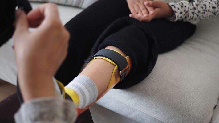
What Is an Orthosis? Why Is an Orthosis Used?
Ankara Ortopedi | Prof. Dr. Murat Demirel » General » What Is an Orthosis? Why [...]
13
Apr
Apr

Knee Replacement Surgery Prices
Ankara Ortopedi | Prof. Dr. Murat Demirel » Knee Treatments » Knee Replacement Surgery PricesThe [...]
18
Aug
Aug
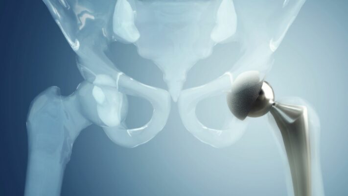
Hip Replacement Surgery Prices
Ankara Ortopedi | Prof. Dr. Murat Demirel » Hip Treatments » Hip Replacement Surgery PricesThe [...]
18
Aug
Aug