

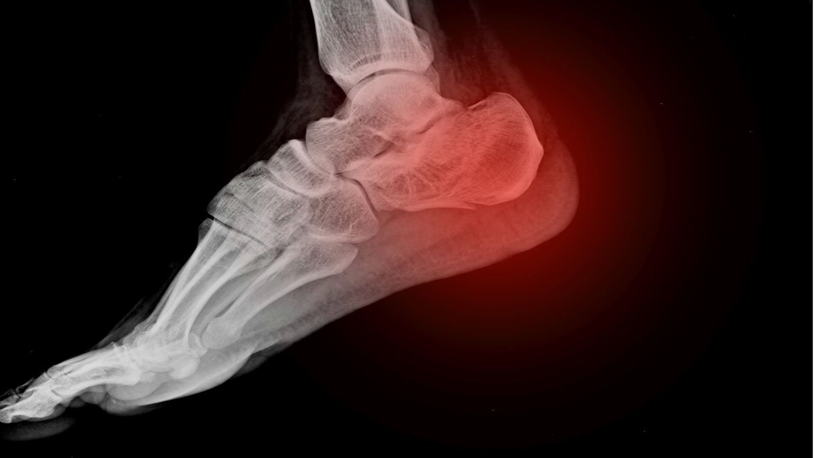
Prof. Dr. Murat Demirel, one of the best orthopedic doctors in Ankara specializing in foot stress fracture treatment, stands out with his long-standing experience in diagnosing and treating these microfractures that occur in the foot bones as a result of repetitive strain and overloading. Foot stress fractures are common, especially among athletes, people who walk extensively, or those who work long hours on hard surfaces, and if left untreated, they may lead to more serious bone injuries. Among the hospitals in Ankara providing foot stress fracture treatment, Prof. Dr. Demirel applies personalized treatment plans to his patients in advanced medical facilities with high hygiene standards.
During the treatment process, depending on the severity of the fracture and the patient’s lifestyle, rest, splint or cast applications, special insoles, physical therapy programs, or surgical methods when necessary are chosen. After foot stress fracture treatment, following the doctor’s recommendations ensures that the healing process is fast, safe, and permanent. In addition, transparent and up-to-date information is provided about foot stress fracture treatment costs in Ankara, helping patients make informed decisions. You can also schedule an appointment immediately to protect your foot health, reduce your pain, and move safely.
| Disease Name | Foot Stress Fracture |
| Affected Area | Metatarsal bones, navicular bone, calcaneus, talus |
| Main Causes | Repetitive and intense loading, insufficient rest, inappropriate footwear, sudden increase in activity |
| Symptoms | Localized foot pain that increases with activity and decreases with rest, swelling, tenderness |
| Risk Factors | Intensive sports activities (running, dancing), osteoporosis, female gender, amenorrhea, malnutrition, training on hard surfaces |
| Diagnostic Methods | Physical examination, X-ray may appear normal initially; definitive diagnosis requires MRI or bone scintigraphy |
| Treatment Methods | Rest, activity modification, ice application, supportive shoes or walking boot; rarely surgery |
| Surgical Necessity | May be required in high-risk areas (navicular, fifth metatarsal base), nonunion or recurrent fractures |
| Physiotherapy Necessity | Generally recommended; to readjust loading and ensure muscle balance |
| Possible Complications | Nonunion, fracture progression, chronic pain, recurrence |
| Recovery Process | Usually 6–8 weeks; full return to sports activities may take 8–12 weeks |
| Follow-Up Necessity | Healing process should be monitored with clinical examination and imaging |


Prof. Dr. Murat Demirel
Orthopedics and Traumatology Specialist
Orthopedics Specialist Prof. Dr. Murat Demirel was born in Ankara in 1974. He completed his primary education at Ankara Kavaklıdere Primary School and his secondary and high school education at Ankara Atatürk Anatolian High School. Dr. Demirel graduated from Ankara University Faculty of Medicine in 1998 and completed his residency in Orthopedics and Traumatology at Ankara Numune Training and Research Hospital, 1st Orthopedics and Traumatology Clinic, in 2004.
PhD
Ankara University Institute of Health Sciences
Specialization
Ankara Numune Training and Research Hospital, 1st Orthopedics Clinic
Medical School
Ankara University Faculty of Medicine
Yazı İçeriği
What Is a Foot Stress Fracture and Why Does It Occur?
The bones in our body are incredibly dynamic and intelligent structures. They constantly renew themselves. This process is called “bone remodeling.” In this cycle, the cells we call “osteoclasts” clean up old and worn bone tissue, while the “osteoblasts” build fresh, new, and strong bone tissue in its place. Every movement we make, every step we take, triggers this cycle. A fundamental principle known as Wolff’s Law states that bone adapts to the loads placed upon it. In other words, our bones function on a “use it or lose it” basis; the more regular and balanced the loading, the stronger they become.
So, how does a foot stress fracture occur within this brilliant system? The problem begins when the balance is disrupted. When the repair team of osteoblasts cannot keep up with the demolition crew of osteoclasts, microscopic damage begins to accumulate in the bone. Imagine a construction site: the demolition team is working too fast, but the builders, due to lack of materials or time, cannot keep up at the same speed. As a result, a weakness or crack develops in the site.
This process generally develops in two main scenarios: fatigue fractures and insufficiency fractures.
Fatigue fractures occur when a completely healthy bone is subjected to excessive and repetitive loading that it is not accustomed to. For example, someone who does not normally exercise deciding to run a marathon, or long and exhausting marches during military training can trigger this. The bone simply does not have the time to adapt to this new and intense pace.
Insufficiency fractures, however, present a different picture. In this case, the problem lies within the bone itself. The bone is already weakened due to osteoporosis, severe vitamin D deficiency, certain medications, or metabolic diseases. Such a weakened bone may break even under daily loads that would normally be easily tolerated (like standing for long periods or shopping). This type of fracture is especially important because it often indicates an underlying and treatable broader health condition. The treatment plan should focus not only on the fracture itself but also on identifying and addressing the cause of the bone weakness.
What Are the Risk Factors in the Development of Foot Stress Fractures?
Foot stress fractures rarely occur due to a single cause. They usually arise from a “perfect storm” of multiple risk factors. Knowing these factors is the most important step in prevention. These risks can be categorized as intrinsic (related to the person) and extrinsic (environmental).
Intrinsic risk factors that are harder to control include:
- Previous stress fracture
- Female gender
- Low bone mineral density
- Menstrual irregularities or absence
- Low body mass index
- High or flat foot arch
- Leg length discrepancy
- Weakness in muscle strength and flexibility
- Genetic predisposition
- Advanced age
Extrinsic risk factors, related to lifestyle and habits, include:
- Sudden increase in training intensity
- Sudden increase in training duration
- Sudden increase in training frequency
- Insufficient rest and recovery
- Training on hard or uneven surfaces
- Worn-out shoes with no support
- Repetitive, monotonous exercises
- Insufficient calorie intake
- Low calcium and vitamin D intake
- Smoking
- Excessive alcohol consumption
Especially in female athletes, a condition known as the “Female Athlete Triad” or, more recently, “Relative Energy Deficiency in Sport (RED-S)” is a serious risk factor for stress fractures. This condition is characterized by insufficient energy intake, menstrual dysfunction, and low bone density, and it requires a professional approach.
What Are the Most Noticeable Symptoms of Foot Stress Fractures?
Symptoms of foot stress fractures usually develop slowly and insidiously. This can delay the recognition of the injury. Most patients cannot give a clear answer to the question “When did it happen?” because there is no sudden trauma. Symptoms worsen gradually over weeks.
The most common signs and symptoms of stress fractures include:
- Pain that increases with activity
- Pain that decreases with rest
- Localized tenderness
- Mild swelling (edema)
- Sometimes redness or bruising
- Night pain (in advanced cases)
- Limping
The nature of the pain is quite typical. Initially, it is felt only during or after strenuous activities like running or training. The person may mistake it for simple muscle soreness and ignore it. However, as the damage progresses, pain appears earlier during activity, intensifies, and eventually even daily activities like walking to the store become painful. In the advanced stage, pain may even occur at rest or while lying in bed at night. Night pain usually indicates significant edema and damage within the bone.
The most important physical examination finding is “point tenderness.” When pressure is applied with a single finger to the painful area, the patient experiences sharp, localized pain, which strongly supports the suspicion of a stress fracture. This tenderness is typically confined to a coin-sized area rather than widespread.
Contact us for detailed information and an appointment!
How Is a Foot Stress Fracture Definitively Diagnosed?
When we suspect a foot stress fracture, we use modern imaging technologies to confirm the diagnosis and plan the correct treatment. After listening to the patient’s detailed history and performing a physical examination, we usually follow these steps.
X-ray: This is usually the first step. It is an easily accessible and quick method. However, due to the nature of stress fractures, X-rays often appear “normal” in the early stages. X-rays can show bone separation or healing tissue (callus), but these findings may take weeks to appear. Therefore, a normal X-ray result while your pain persists should not mislead you; it does not mean that you do not have a stress fracture, only that it has not yet progressed enough to be visible on X-ray.
Magnetic Resonance Imaging (MRI): MRI is the gold standard in diagnosing stress fractures. An MRI image of a stress fracture can show even the smallest changes inside the bone. It can clearly detect bone marrow edema (a type of internal bruising) even in the “stress reaction” phase before a crack develops. This allows us to diagnose very early and start treatment immediately, preventing progression to a full fracture. MRI also evaluates other soft tissues in the foot (muscles, tendons, ligaments) to help determine whether the pain is caused by another condition.
Computed Tomography (CT): CT is a method that shows bone structure in three dimensions and in great detail. It can be used especially in stress fractures that are difficult to heal and classified as “high risk” (such as the navicular bone), or when planning surgery. It provides valuable information on the exact location of the fracture, whether there is displacement, and monitoring the healing process.
Bone Scintigraphy: This method shows bone activity throughout the skeletal system. It is highly sensitive in detecting stress fractures but has low specificity. In other words, it may show positive results in conditions other than fractures, such as infections, arthritis, or tumors. With the widespread use of MRI, its role in diagnosing stress fractures has significantly decreased.
Why Are Foot Stress Fractures Divided Into Different Categories?
We cannot treat all stress fractures the same way. Each bone has a different anatomical location, blood supply, and load distribution. The most important roadmap in determining treatment strategy is to classify fractures as “low risk” or “high risk.” This distinction is vital for the patient’s healing process and future.
Foot stress fractures in low-risk areas usually have good blood circulation, are exposed to compressive forces, and therefore have high healing potential. These are usually treated successfully with non-surgical methods.
Some of these areas include:
- Calcaneus (heel bone)
- Shafts of the metatarsal bones (especially 2nd, 3rd, and 4th metatarsals)
- Cuboid and cuneiform bones
In contrast, stress fractures in high-risk areas often occur in zones with poor blood supply (“avascular” areas) and are subjected to continuous tension or twisting forces. Therefore, delayed healing (delayed union) or no healing (nonunion) is a serious risk. These cases require more aggressive treatment and sometimes direct surgical intervention.
The most well-known high-risk areas include:
- Navicular bone (a key bone in the midfoot with poor blood supply)
- Proximal fifth metatarsal (the area near the base of the little toe, also known as a “Jones fracture”)
- Talus bone (forms the ankle joint)
- Medial malleolus (the inner ankle prominence)
- Sesamoid bones (small round bones under the big toe)
Making this classification determines whether we say “rest for a few weeks, it will heal” or “you need a 6-week cast with no weight-bearing, or even surgery.”
What Are the Non-Surgical Treatment Methods for Foot Stress Fractures?
The good news is that most foot stress fractures, especially low-risk ones, heal completely without the need for surgery, with the right conservative treatment plan. The basic philosophy of this treatment is simple: give the fractured bone the peace and time it needs to heal.
The main components of non-surgical treatment include:
- Activity modification
- Load protection (immobilization)
- Ice application
- Elevation (keeping the foot elevated)
- Nutritional support
- Physical therapy
The first and most important step in treatment is to completely stop the activity causing the pain. This is called “relative rest.” The duration varies from 4 to 8 weeks depending on the type of fracture. To maintain conditioning during this period, sports that do not load the foot such as swimming, aqua jogging, or cycling may be done.
Load protection is critical to reducing stress on the fracture site. Depending on the severity and location of the fracture, various methods are used:
- Crutches
- Stiff-soled shoes
- Walking boots (such as Aircast)
- Cast
In a high-risk navicular fracture, a cast with no weight-bearing for 6–8 weeks is required, while in a low-risk metatarsal fracture, controlled weight-bearing with a special walking boot may be permitted.
To control pain and swelling, regular ice application in the first few days and keeping the foot elevated above heart level are very useful. Bone healing is like a construction project and requires the right “building materials.” Therefore, a diet rich in calcium and vitamin D, along with supplements if necessary, supports the healing process.
It is important to warn patients about methods such as “herbal treatment for stress fractures.” Relying on such scientifically unproven methods and delaying modern medical treatment can increase the risk of nonunion—the most common cause of “stress fracture not healing” complaints—and lead to much more complicated treatments.
When Is Surgery an Option for Foot Stress Fractures?
Surgery is a less frequent option in the treatment of foot stress fractures, but in some cases it is unavoidable and the most appropriate choice. The decision for surgery depends on several important factors.
Main situations that may require surgical intervention include:
- Fractures in high-risk areas
- Failure of non-surgical treatment (nonunion)
- Displacement of fracture fragments
- Elite athletes aiming for rapid return to sports
Especially in fractures of high-risk areas such as the navicular or the 5th metatarsal (Jones fracture), where blood supply is poor, choosing surgery initially may provide a more predictable and reliable outcome compared to prolonged non-surgical treatment. Non-healing fractures, even after months of rest and casting, are also treated surgically.
The goal of surgical technique is to stabilize the fracture fragments tightly, preventing movement and creating an ideal environment for union. This is usually referred to as “internal fixation.”
Some surgical materials used include:
- Screws
- Plates
- Wires
For example, in a Jones fracture, a single screw placed inside the bone can perfectly stabilize the fracture line. In cases of nonunion or poor biological healing, bone grafting (using a small piece of bone usually taken from the hip) or bone marrow concentrate (stem cells) may be added to biologically support healing. Surgery is also valuable for elite athletes because it offers a clearer and often faster timeline for returning to sports compared to an uncertain and prolonged non-surgical healing process.
How Does the Recovery and Rehabilitation Process Progress After a Foot Stress Fracture?
Bone healing is only the first half of treatment. The real goal is for the patient to safely and fully return to daily life and sports. This is where rehabilitation, including physical therapy and exercise programs, comes into play. Rehabilitation is not just waiting for bone union, but regaining all the functional losses caused by the injury.
The main goals of rehabilitation are:
- Control of pain and swelling
- Restoration of joint range of motion
- Increase in muscle strength
- Improvement of flexibility
- Enhancement of balance and coordination (proprioception)
- Relearning correct movement patterns
Rehabilitation generally progresses with a phased protocol. In the initial phase, while pain and swelling are controlled, passive and active range of motion exercises are performed to prevent joint stiffness. When full weight-bearing without pain is possible, strengthening exercises are introduced. Strengthening the ankle, calf, knee, and hip muscles helps absorb loads placed on the foot in the future.
Balance and coordination exercises improve the body’s ability to perceive its position in space, reducing the risk of reinjury. In the final stage, sport- or activity-specific movements (running, jumping, changing direction) are gradually added to the program. Rushing the return to sports is dangerous. The answer to “When can I start running?” depends not on the calendar but on the patient’s functional condition. Pain is the most important guide. Even the slightest pain during activity indicates that the body is not ready for that level yet.
What Can Be Done to Prevent Foot Stress Fractures in the Future?
Experiencing a stress fracture once provides valuable lessons for preventing recurrence in the future. Prevention is always easier and more important than treatment. A multi-faceted approach is necessary to prevent foot stress fractures.
Training-related precautions include:
- Gradual progression (“10% rule”)
- Sufficient rest days
- Cross-training (engaging in different sports)
- Proper warm-up and cool-down routines
- Variety in training surfaces
Equipment and footwear selection also play a critical role:
- Sport-specific shoes
- Good cushioning and support
- Regular replacement of worn shoes (every 600–800 km on average)
- Custom orthotics if necessary
Nutrition is the foundation of bone health:
- Adequate calorie intake
- Calcium-rich foods (dairy products, leafy greens)
- Sufficient vitamin D (sunlight and supplements if necessary)
- Adequate protein intake
- Magnesium and vitamin K
Finally, learning to listen to the signals your body sends is perhaps the most important preventive method. Pain is not an enemy but a friend. It alerts you when something is wrong. The culture of “pushing through pain,” common among athletes, is the biggest invitation to overuse injuries like stress fractures. Take care of your body, give it the rest and nutrition it needs, and it will not let you down.
Contact us for detailed information and an appointment!
Frequently Asked Questions
What is a foot stress fracture and how does it occur?
A foot stress fracture is a small, incomplete crack in the long bones of the foot or metatarsals, caused by repetitive minor trauma, overuse, or excessive strain. It is most commonly seen in long-distance runners, soldiers, dancers, or people with a sudden increase in activity. Inappropriate footwear and weakened bone structure are also risk factors.
What are the symptoms of a stress fracture?
Initially, there may be mild pain and tenderness, but over time swelling, pain when bearing weight, and slight redness of the foot may develop. The pain typically decreases with rest but returns with activity. There is usually no history of significant trauma.
How is a stress fracture diagnosed?
On physical examination, there is tenderness, swelling, and pain over the bone. In the early stages, X-rays may not show any abnormalities; therefore, advanced imaging methods such as magnetic resonance imaging (MRI) or bone scintigraphy are often required for a definitive diagnosis.
How is a foot stress fracture treated?
The basis of treatment is rest and reducing the load on the foot. Activity is restricted, and crutches or custom insoles may be used if necessary. Ice can be applied to the painful area, and pain relievers may be recommended. Depending on the location and severity of the fracture, short-term casting or splinting may be needed.
Does a stress fracture require surgery?
Most stress fractures heal without surgery. However, if the fracture does not heal, does not unite, or if the bone fragments are displaced, surgical intervention may be considered. Surgery is generally rarely required.
How long does the recovery process take?
Complete healing of stress fractures usually takes 6–8 weeks. During this period, activities should be gradually increased, and overloading should be avoided. Returning to sports or heavy activities too early is not recommended.
Can a stress fracture recur?
If the underlying causes (overuse, bone weakness, inappropriate footwear) are not corrected, stress fractures can recur. Ensuring bone strength and taking preventive measures are necessary to avoid recurrence.
What should people with stress fractures pay attention to?
Adequate rest, healthy and balanced nutrition, sufficient intake of vitamin D and calcium, choosing proper footwear for foot health, and gradually increasing weight-bearing activities are important.
How should return to sports be managed after a stress fracture?
Sports should not be resumed until full recovery is achieved. Activities should not be started without the approval of a doctor and physiotherapist. When returning to sports, load should be increased gradually.
Can stress fractures be prevented?
Gradual and controlled increases in activity, proper training techniques, appropriate footwear selection, regular rest intervals, and maintaining bone health are effective in preventing stress fractures.
Blog Posts

What Is a Tendon Rupture? Symptoms, Causes, and Treatment Methods
Ankara Ortopedi | Prof. Dr. Murat Demirel » General » What Is a Tendon Rupture? [...]
13
Apr
Apr

What Is an Orthosis? Why Is an Orthosis Used?
Ankara Ortopedi | Prof. Dr. Murat Demirel » General » What Is an Orthosis? Why [...]
13
Apr
Apr

Knee Replacement Surgery Prices
Ankara Ortopedi | Prof. Dr. Murat Demirel » Knee Treatments » Knee Replacement Surgery PricesThe [...]
18
Aug
Aug
Hip Replacement Surgery Prices
Ankara Ortopedi | Prof. Dr. Murat Demirel » Hip Treatments » Hip Replacement Surgery PricesThe [...]
18
Aug
Aug