

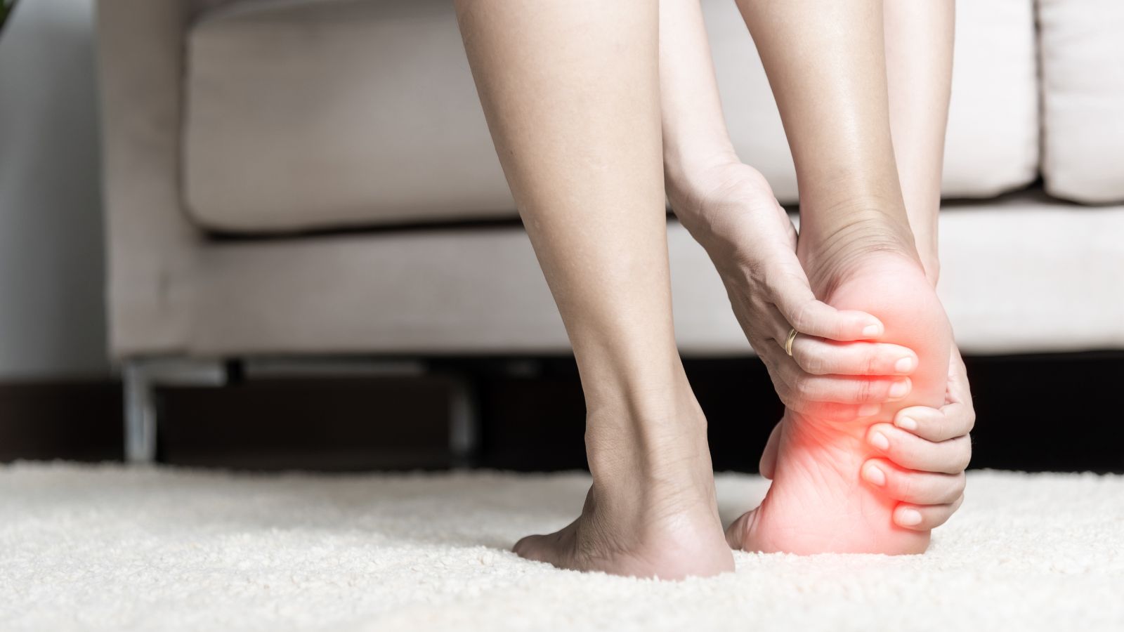
Prof. Dr. Murat Demirel, one of the best orthopedic doctors in Ankara for the treatment of talus osteochondral lesions (cartilage damage), stands out with his many years of experience in diagnosing and treating damage to the cartilage and underlying bone tissue in the ankle joint. Talus osteochondral lesions usually occur as a result of ankle sprains, traumas, or repetitive strains, and over time can cause pain, swelling, and limitation of movement in the joint. Among the hospitals in Ankara that treat talus osteochondral lesions, Prof. Dr. Demirel offers personalized treatment plans to his patients in centers with advanced medical equipment and high hygiene standards.
Depending on the severity of the damage, treatment options may include rest, physical therapy, PRP or stem cell injections, microfracture technique, cartilage transplantation, or arthroscopic surgical techniques. After talus osteochondral lesion treatment, adherence to the doctor’s recommendations ensures a fast, safe, and lasting recovery. In addition, transparent and up-to-date information about the cost of talus osteochondral lesion treatment in Ankara is provided, helping patients make informed decisions. You can also protect your ankle health, reduce pain, and increase mobility by contacting us to book an appointment right away.
| Disease Name | Talus Osteochondral Lesions (Cartilage Damage) |
| Affected Area | Talus bone (cartilage-covered area located in the ankle joint) |
| Main Causes | Traumatic ankle sprains, repetitive microtraumas, circulatory disorders |
| Symptoms | Deep, localized pain in the ankle; difficulty walking, catching sensation, swelling, limitation of movement |
| Risk Factors | Frequent sprains in athletes, anatomical abnormalities, inadequately treated ankle traumas |
| Diagnostic Methods | MRI (most sensitive method for location and depth of lesion), CT, X-ray; sometimes arthroscopy |
| Treatment Methods | Conservative: rest, load reduction, physical therapy; Surgical: arthroscopic debridement, microfracture, mosaicplasty, osteochondral autograft, or autologous chondrocyte implantation |
| Surgical Necessity | Applied in symptomatic and deep lesions unresponsive to conservative treatment |
| Physiotherapy Requirement | Recommended in both conservative and post-surgical recovery; necessary to maintain range of motion and prepare for loading |
| Possible Complications | Insufficient cartilage healing, recurrence, development of osteoarthritis, joint stiffness, persistent pain |
| Recovery Process | Varies depending on the treatment method, but after surgery 6–12 weeks, full functional recovery may take months |
| Follow-up Requirement | Requires clinical check-ups, MRI follow-ups, and monitoring throughout the rehabilitation process |

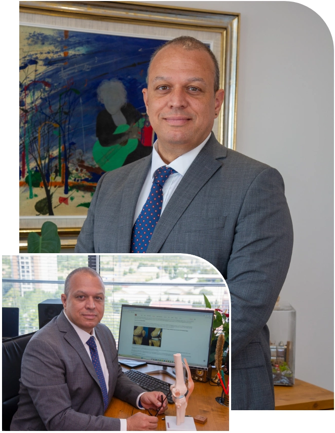
Prof. Dr. Murat Demirel
Orthopedics and Traumatology Specialist
Orthopedics Specialist Prof. Dr. Murat Demirel was born in Ankara in 1974. He completed his primary education at Ankara Kavaklıdere Primary School and his secondary and high school education at Ankara Atatürk Anatolian High School. Dr. Demirel graduated from Ankara University Faculty of Medicine in 1998 and completed his residency in Orthopedics and Traumatology at Ankara Numune Training and Research Hospital, 1st Orthopedics and Traumatology Clinic, in 2004.
PhD
Ankara University Institute of Health Sciences
Specialization
Ankara Numune Training and Research Hospital, 1st Orthopedics Clinic
Medical School
Ankara University Faculty of Medicine
Yazı İçeriği
What are talus osteochondral lesions and how do they affect the ankle?
This term may sound complicated at first, so let’s simplify it. The answer to the question “What is an osteochondral lesion?” lies in two words: “Osteo” means bone, “chondral” means cartilage. So, an osteochondral lesion refers to damage that affects both the joint cartilage and the bone tissue immediately beneath it. At the center of the ankle joint, between the leg bones (tibia and fibula) and the heel bone (calcaneus), there is a bone called the talus that functions like a hinge. The entire weight of our body is transmitted to the foot through this small bone. The articular surface of this bone is covered with a smooth, shiny, and slippery layer of cartilage that eliminates friction. This cartilage is a perfect design that allows the joint to move millions of times painlessly and without catching.
When a talus osteochondral lesion occurs, this perfect design is disrupted. The damage can be a superficial crack in the cartilage (chondral lesion), or it may progress into a more serious condition where a piece of bone under the cartilage separates along with it (osteochondral lesion). Sometimes the cartilage surface may appear intact, while the actual problem is in the underlying bone, manifesting as collapse or cyst formation due to impaired blood supply (subchondral lesion).
The effect of this damage on ankle function is like a chain reaction. When the smooth surface deteriorates, friction increases, which directly causes pain. The body perceives this abnormal situation as a threat and produces more joint fluid to protect the joint, leading to swelling. If there is a free-floating cartilage or bone fragment, it can get caught between joint movements, causing mechanical issues such as catching, locking, and sudden pain attacks. Most importantly, over time this damaged surface disrupts the balance of the entire joint, paving the way for ankle osteoarthritis, an irreversible condition.
Why do talus osteochondral lesions occur?
The most common scenario in the development of these lesions is overwhelmingly trauma. A severe ankle sprain is the number one cause of this damage. During the sprain, the talus bone is compressed like in a vise by the other bones forming the joint. This sudden and severe pressure can lead to crushing or fracture of the sensitive cartilage surface and underlying bone. Studies show that more than half of patients with ankle sprains and nearly three-quarters of those with ankle fractures have varying degrees of cartilage damage.
However, not every lesion has a clear history of trauma. In some cases, especially lesions seen on the inner side of the talus, they may develop without significant trauma. This condition is called “osteochondritis dissecans.” In this scenario, the problem is usually impaired blood circulation in the bone beneath the cartilage. By nature, the blood supply to the talus bone is already somewhat weak. Repeated small, unnoticed impacts (such as microtraumas in certain athletes) or sometimes completely unknown reasons prevent enough blood from reaching this bone area. The poorly nourished bone tissue weakens, loses vitality, and eventually collapses. This process, also known as “talus bone necrosis” among the public, leads to cartilage damage over time as the weakened base fails to support it.
What are the most common symptoms of talus osteochondral lesions?
Talus osteochondral lesions can sometimes be insidious and initially produce very few symptoms. However, in most patients, especially when the lesion reaches a certain size or becomes unstable, typical complaints arise. The most common symptoms we encounter are:
- Deep and aching joint pain
- Pain that increases especially with weight bearing (walking, running)
- Visible swelling in the joint
- Restricted ankle movement
- Feeling of stiffness in the joint
- Sensation of catching or “jumping”
- Locking (joint getting stuck in one position)
- Feeling of instability or “giving way” in the ankle
The most important clue for patients here is the condition described as an “ankle sprain that does not heal.” If you have experienced a sprain and weeks have passed without completing the normal healing process—persistent pain and swelling remain—then a cartilage injury must be suspected. This is a red flag situation requiring advanced evaluation when standard treatments fail.
Contact us for detailed information and an appointment!
How are talus osteochondral lesions diagnosed?
Accurate diagnosis is the foundation of proper treatment. Therefore, we manage the diagnostic process meticulously, step by step. The first step is always to listen carefully to the patient. The history of your complaints, when and how they began, and whether you have experienced trauma before provide the first clues for diagnosis. Then, with a comprehensive physical examination, we evaluate the condition of your ankle, the strength of your ligaments, and sensitive points.
However, imaging methods are indispensable for definitive diagnosis and mapping the lesion in detail. The main tools we use in this process and what each tells us are as follows:
X-ray: Usually the first step. It provides a general idea about bone structures. But it must be remembered that X-rays do not show cartilage. Therefore, especially in early-stage lesions or cases with only cartilage damage, X-ray images may appear completely normal. This does not mean that the patient has no pain.
Magnetic Resonance Imaging (MRI): Considered the “gold standard” in diagnosing talus osteochondral lesions. MRI provides us with very valuable and detailed information about the lesion. This includes:
- Exact location, size, and depth of cartilage damage
- Whether there is edema (bone marrow injury) in the bone beneath the cartilage
- Presence of an underlying cyst
- Whether the lesion is stable (in place) or unstable (at risk of separation)
- Whether there is a free fragment (loose body) in the joint
Computed Tomography (CT): Used especially when a three-dimensional and very detailed examination of bone structures is required. Particularly if surgery is planned, CT acts as a roadmap to clearly reveal the exact size of the bone damage, the structure of cysts, and the position of detached fragments.
These imaging methods are complementary, not alternatives to each other. Which tests are necessary is decided based on your specific condition, complaints, and examination findings.
How are talus osteochondral lesions classified according to severity?
After diagnosing the lesion, the next step is to determine its severity and character. To do this, we rely on imaging findings (especially MRI) and sometimes arthroscopic observations (looking directly into the joint during minimally invasive surgery). This staging guides us in selecting the most appropriate treatment method. To make it easier for our patients to understand, we can explain this classification like a story:
Everything begins with a “bruise” or “bone marrow edema” beneath the cartilage (Stage 1). At this stage, the cartilage surface is still intact, but a problem has started at its foundation. If this pressure continues, a “crack” forms in the cartilage and bone above (Stage 2). This crack partially separates the fragment, but part of it is still attached to the main bone.
If the process advances, this fragment “completely breaks off” but still remains in its bed without displacing (Stage 3). This is like a broken but not scattered vase. In the final stage, this detached fragment separates completely from its bed and “falls into the joint space” (Stage 4). At this point, it becomes a free “loose body” in the joint, with the potential to cause problems with every movement. Sometimes instead of these stages, a fluid-filled “cyst” may also form inside the bone over time (Stage 5).
What are the non-surgical treatment options for talus osteochondral lesions?
Indeed, not every talus osteochondral lesion requires immediate surgery. Especially in early-stage, small, and stable (non-displaced) lesions, it is possible to achieve successful results with non-surgical (conservative) treatments. The basic philosophy of this treatment is to eliminate stress on the damaged area and allow the body’s self-healing potential to take effect. This approach is more commonly preferred in children and young people, as their bone and cartilage healing capacity is higher.
Our non-surgical treatment protocol generally includes the following steps:
Activity Modification: Complete avoidance of activities that trigger pain such as running, jumping, and strenuous exercise.
Non-weight Bearing (Immobilization): This is the most critical part of the treatment. Typically, no weight is placed on the affected leg for 4 to 6 weeks, using crutches. During this period, the ankle may be immobilized with a special walking boot or sometimes a cast.
Physical Therapy and Rehabilitation: After the non-weight-bearing period ends, a gradual program begins under the supervision of a physiotherapist. The goals of this program are:
- To regain joint range of motion
- To strengthen the muscles around the ankle
- To improve balance and proprioception (sense of position)
- To restore a normal and pain-free walking pattern
Medication: Non-steroidal anti-inflammatory drugs may be prescribed by the doctor to control pain and inflammation.
Biological Injections: In recent years, intra-articular injections such as PRP (Platelet-Rich Plasma) or hyaluronic acid have also been included in treatment options to support the healing environment. These methods may help relieve symptoms.
The success rate of non-surgical treatment varies depending on the type of lesion and patient compliance, but in adults, it is around 50%. If after a trial period of 3–6 months there is no significant improvement in complaints or if symptoms worsen, surgical treatment is considered the next step.
When is surgery necessary for talus osteochondral lesions?
The decision for surgery is an important one, made after evaluating the patient’s condition from multiple aspects. The answer to “When is surgery necessary?” depends on several key factors. The most common situations leading us to consider surgical treatment are:
- Failure of non-surgical treatment after 3 to 6 months.
- Mechanical symptoms such as catching or locking in the ankle.
- MRI or other imaging showing that the lesion is unstable (mobile).
- Presence of a displaced fragment in the lesion.
- Presence of a loose cartilage or osteochondral fragment (“loose body”) in the joint.
- Lesions above a certain size (generally larger than 1–1.5 cm²).
- Presence of a large bone cyst beneath the lesion.
- Young, active patients with high expectations of returning to sports (if lesion features are suitable).
Especially mechanical symptoms and instability seen on imaging usually require direct surgery. Because the likelihood of spontaneous healing in such cases is very low, and they have a high potential to cause further joint damage.
What surgical methods are used for talus osteochondral lesions?
Talus cartilage surgery, or more accurately TOL surgery, is like tailoring a custom suit; there is no single standard method. The surgical technique is carefully chosen based on the size, stage, location of the lesion, and the patient’s individual characteristics. Thanks to advances in technology, most of these surgeries can now be performed arthroscopically (minimally invasive), meaning through just a few small incisions. This results in less pain, faster recovery, and better cosmetic outcomes.
Our main goals in surgical treatment are:
- To eliminate pain and restore function.
- To repair or regenerate the damaged cartilage tissue.
- To restore the joint’s normal mechanics and surface alignment.
- To minimize the risk of future osteoarthritis.
The main surgical methods we use to achieve these goals are:
Debridement and Bone Marrow Stimulation Techniques (Microfracture): Usually our first choice for small (less than 1.5 cm²), non-cystic lesions. First, the damaged and loose cartilage fragments are arthroscopically cleaned (debridement). Then, small holes are made in the bone at the base of the lesion using a special tool, creating controlled bleeding (microfracture). The stem cells from the bone marrow in this blood form a clot in the damaged area, which later turns into a repair tissue called “fibrocartilage.” Although this new tissue is not as durable as the original cartilage, it effectively fills the defect, reduces pain, and improves function.
Osteochondral Autograft Transfer (OATS / Mosaicplasty): Applied in larger lesions or when microfracture treatment is insufficient. This technique involves transplanting bone-cartilage plugs with healthy original cartilage from a non-weight-bearing area of the patient’s knee joint. These cylindrical plugs are then implanted into the damaged talus area in a mosaic pattern. The biggest advantage of this method is directly replacing the damaged area with durable, original-like hyaline cartilage.
Autologous Chondrocyte Implantation (ACI / MACI): Usually reserved for large lesions, this is an advanced, two-step cell therapy technique. In the first stage, a small healthy cartilage sample is arthroscopically taken from the patient’s knee. This sample is then cultured in a special laboratory for 3–4 weeks to produce millions of new cartilage cells. In the second surgery, these laboratory-grown cells are implanted into the cleaned lesion site on the talus. The goal is to create high-quality repair tissue that closely resembles original hyaline cartilage.
Fragment Fixation and Bone Grafting: If the lesion results from a large, viable osteochondral fragment broken off by trauma, instead of removing it, we can reattach it to its original bed using special absorbable screws, pins, or sutures (fixation). If there is a large cyst beneath the lesion, we can clean it out and fill it with bone graft from the patient or synthetic bone substitutes.
What is the recovery process after surgery for talus osteochondral lesions?
Just as important as the success of the surgery itself is the postoperative rehabilitation process, which is the most crucial factor determining the final outcome. This process is like running a marathon; it requires patience, discipline, and care. Frequently asked “ankle cartilage surgery patient reviews” also emphasize how important this process is. The recovery process varies slightly depending on the surgery performed, but generally follows certain phases:
Phase 1: Maximum Protection Phase (First 6–8 weeks after surgery): This is the most critical period when the newly repaired tissue is at its most vulnerable and must be protected. The main rules in this phase are:
- Absolutely no weight bearing
- Use of crutches
- Protecting the ankle with a special boot or brace
- Regular ice application and elevating the leg to control swelling
- Starting gentle, passive movements as instructed by your doctor and physiotherapist
Phase 2: Gradual Weight Bearing and Regaining Motion (Usually weeks 8–12): With your doctor’s approval, you begin to gradually and carefully put weight on your ankle. Physiotherapy sessions intensify during this period. The goal is to fully restore joint mobility and relearn a normal walking pattern.
Phase 3: Strengthening and Return to Function (Usually 3–6 months): At this stage, exercises aimed at strengthening the muscles around the ankle, and improving balance and coordination come to the forefront. Low-impact cardio such as stationary cycling may be started. Return to daily activities is usually completed in this phase.
Phase 4: Return to Sports (Usually 6–12 months and beyond): This final stage requires the most patience. Returning to demanding and sport-specific activities such as running and jumping is done gradually, only after passing certain strength and functional tests. Full return to sports may take up to a year depending on the type of surgery, the level of sport, and the individual’s healing speed.
Contact us for detailed information and an appointment!
Frequently Asked Questions
What is a talus osteochondral lesion and how does it occur?
Talus osteochondral lesions occur when the cartilage and the underlying bone tissue located on the surface of the talus bone in the ankle joint are damaged. They usually result from an ankle sprain, a fall, sports injuries, or repetitive microtraumas. Sometimes circulatory disorders or spontaneous weakening of the cartilage may also play a role.
What are the symptoms of talus cartilage damage?
Deep and persistent ankle pain, restricted movement, swelling in the joint, sometimes a catching or locking sensation, pain that increases during long walks, and occasionally clicking sounds from the joint are the main symptoms. Complaints usually start with persistent pain following a sprain.
How are talus osteochondral lesions diagnosed?
Diagnosis involves patient history, physical examination, and radiological imaging. Standard X-rays may sometimes be insufficient, and for definitive diagnosis magnetic resonance imaging (MRI) is usually required. MRI provides detailed information about the size and location of cartilage and bone damage.
Can cartilage damage heal on its own?
Since cartilage tissue is avascular, it is difficult for it to heal completely on its own. In small and superficial lesions, symptoms may improve, but in larger and deeper lesions, treatment is necessary. If left untreated, pain and movement restrictions may become permanent.
What treatment methods are used for talus osteochondral lesions?
In mild cases, rest, reducing load, physical therapy, painkillers, and special insoles are recommended. In moderate and advanced lesions, arthroscopic surgery can be performed to clean damaged cartilage, with cartilage repair procedures such as microfracture or mosaicplasty. In some patients, stem cell therapy or cartilage transplantation may also be considered.
What is the recovery process after treatment?
After surgery, patients generally avoid full weight bearing for the first few weeks and use crutches. With physical therapy and gradual loading, joint mobility and muscle strength improve. Full recovery and return to sports may take 3–6 months, depending on the severity of the damage.
Do talus osteochondral lesions recur?
With proper treatment and rehabilitation, the risk of recurrence decreases. However, if trauma recurs, excessive loading occurs, or ankle instability persists, lesions may reappear.
What can be done to prevent cartilage damage?
Strengthening the ankle muscles, wearing appropriate shoes, warming up adequately before sports, taking precautions to reduce the risk of sprains, and avoiding repetitive trauma are effective in protecting cartilage health.
What happens if talus osteochondral lesions are left untreated?
Untreated cartilage damage can gradually progress, leading to chronic joint pain, restricted movement, and early-onset ankle osteoarthritis. This can significantly reduce quality of life.
Is it possible to return to sports after ankle cartilage damage?
After successful treatment and rehabilitation, most patients can return to daily activities and light sports. However, starting sports before full recovery may cause the cartilage damage to recur, so returning to sports must always be under a doctor’s supervision.
Blog Posts

What Is a Tendon Rupture? Symptoms, Causes, and Treatment Methods
Ankara Ortopedi | Prof. Dr. Murat Demirel » General » What Is a Tendon Rupture? [...]
13
Apr
Apr
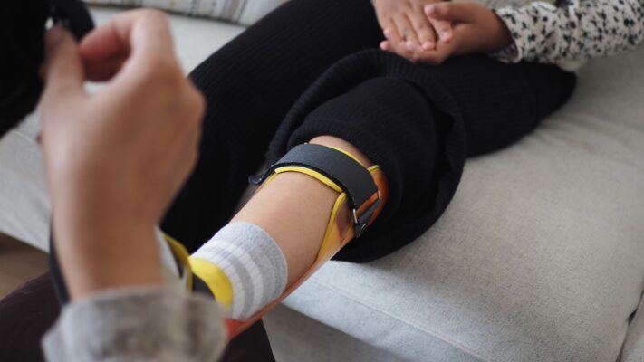
What Is an Orthosis? Why Is an Orthosis Used?
Ankara Ortopedi | Prof. Dr. Murat Demirel » General » What Is an Orthosis? Why [...]
13
Apr
Apr

Knee Replacement Surgery Prices
Ankara Ortopedi | Prof. Dr. Murat Demirel » Knee Treatments » Knee Replacement Surgery PricesThe [...]
18
Aug
Aug
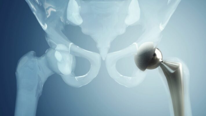
Hip Replacement Surgery Prices
Ankara Ortopedi | Prof. Dr. Murat Demirel » Hip Treatments » Hip Replacement Surgery PricesThe [...]
18
Aug
Aug