


Prof. Dr. Murat Demirel, one of the best orthopedic doctors in Ankara specializing in ankle fracture treatment, stands out with his many years of experience in the diagnosis and treatment of these serious joint injuries caused by trauma, falls, or sports injuries. If not treated correctly, ankle fractures can lead to joint deformity, restricted mobility, and permanent pain. Prof. Dr. Demirel provides his patients with personalized surgical or non-surgical treatment plans in hospitals in Ankara equipped with advanced medical technology and high hygiene standards.
During the treatment process, depending on the type of fracture and the patient’s overall health condition, non-surgical methods such as casts or splints, or surgical options such as fixation with plates and screws, as well as modern rehabilitation programs, are chosen. Following ankle fracture treatment, doctor’s recommendations ensure that the healing process is fast, safe, and permanent. In addition, transparent and up-to-date information about ankle fracture treatment prices in Ankara is provided, helping patients make informed decisions. To protect your ankle health, regain your mobility, and step into a pain-free life, you can contact us now to make an appointment.
| Disease Name | Ankle Fracture |
| Affected Area | Ankle (tibia, fibula, and talus bones) |
| Main Causes | Trauma (fall, sprain, traffic accident), sports injuries, falling from height |
| Symptoms | Severe pain, swelling, bruising, deformity, inability to bear weight, restricted movement |
| Risk Factors | Osteoporosis, advanced age, sports activities, coordination disorders |
| Diagnostic Methods | Physical examination, X-ray, if necessary CT and MRI |
| Treatment Methods | Conservative (cast, orthosis, rest) or surgical (screws, plates) |
| Need for Surgery | Applied in open fractures, those affecting the joint surface, displaced or unstable fractures |
| Need for Physical Therapy | Recommended after casting or surgery to restore joint mobility and muscle strength |
| Possible Complications | Joint stiffness, nonunion, malunion, infection, vascular-nerve injury, development of arthritis |
| Healing Process | 6–12 weeks depending on the location and treatment method of the fracture; full return to activity may take several months |
| Need for Follow-up | The healing process should be monitored with clinical and radiological controls |


Prof. Dr. Murat Demirel
Orthopedics and Traumatology Specialist
Orthopedics Specialist Prof. Dr. Murat Demirel was born in Ankara in 1974. He completed his primary education at Ankara Kavaklıdere Primary School and his secondary and high school education at Ankara Atatürk Anatolian High School. Dr. Demirel graduated from Ankara University Faculty of Medicine in 1998 and completed his residency in Orthopedics and Traumatology at Ankara Numune Training and Research Hospital, 1st Orthopedics and Traumatology Clinic, in 2004.
PhD
Ankara University Institute of Health Sciences
Specialization
Ankara Numune Training and Research Hospital, 1st Orthopedics Clinic
Medical School
Ankara University Faculty of Medicine
Yazı İçeriği
Why Are Ankle Fractures Important and Which Bones Do They Involve?
To understand the seriousness of ankle fractures, one must first grasp the perfect structure of this joint. The ankle is much more than a simple hinge; it is a complex and dynamic structure formed by three bones and the strong ligaments that hold them together. We can compare this structure to the foundation of a house; if the foundation is solid, the building stands firm.
This foundation is made up of three main bones. The tibia (shin bone), which is the main supporting column on the inner side of the leg, forms the medial malleolus, the bony prominence on the inside of the ankle. Next to the tibia is the fibula, a thinner bone on the outer side of the leg that plays a balancing role. The lower end of the fibula forms the lateral malleolus, the bony prominence on the outer ankle. Between these two bones sits the talus, which acts like a ball in a socket, transferring forces between the leg and the foot.
However, this bony framework alone means nothing. There are very strong ligaments that hold them together, provide stability to the joint, and act like steel cables of a building. Especially the syndesmosis, the ligament complex binding the tibia and fibula tightly together, is vital for ankle integrity. When a fracture occurs, these “steel cables” are often strained or completely torn. Therefore, ankle fractures should not be seen as only a bone injury but as a complex event that disrupts the entire balance and mechanics of the joint. Correct diagnosis and treatment of ligament damage is just as important as the fracture itself and directly affects long-term success.
How Do Ankle Fractures Usually Occur?
Ankle fractures usually occur when the joint suddenly and unexpectedly encounters a force greater than it can withstand. These can be quite ordinary scenarios encountered in daily life.
Twisting and Sprains: The most common cause. A missed curb, a slipping rug, a hole in the garden, or a misstep on the stairs can cause the ankle to twist abnormally inward or outward. This sudden and forceful twist acts like a lever on the bones, leading to fractures. Many incidents commonly referred to as “a bad sprain” are actually ankle fractures.
Falls: Jumping from a height, slipping on ice, or even falling awkwardly from a chair can suddenly load the entire body weight on a single ankle. This vertical force often causes more serious fractures (pilon fractures) where the talus embeds into the tibia.
Direct Blows and Accidents: Traffic accidents, a heavy object falling on the foot, or a direct kick during sports can cause complex ankle fractures, often breaking bones in multiple places and into fragments.
Sports Injuries: Sports such as football, basketball, volleyball, skiing, and tennis, which involve sudden stops, accelerations, and directional changes, pose a high risk for ankle fractures. Collisions with opponents or awkward landings after jumps are common injury mechanisms.
Which Conditions Increase the Risk of Ankle Fractures?
Certain personal and environmental factors can make an individual more prone to ankle fractures. Knowing these risk factors is important for prevention. The main risk factors are:
- Osteoporosis
- Low bone density (osteopenia)
- Previous poorly treated ankle sprains
- Active participation in contact and high-impact sports
- Wearing inappropriate footwear that does not support the foot structure
- Deficiency of critical vitamins and minerals for bone health such as vitamin D and calcium
- Balance problems due to advanced age or neurological diseases
- Smoking, which seriously impairs bone circulation and healing capacity
What Are the Symptoms of Ankle Fractures?
When an ankle fracture occurs, your body usually sends such clear and severe signals that it is impossible to ignore. These symptoms are alarm bells to understand the seriousness of the injury. Typical ankle fracture symptoms include:
- Sharp, throbbing pain that starts at the time of injury and worsens
- Significant swelling around the ankle within minutes
- Bruising that appears and spreads within hours or a day
- Inability to bear even the slightest weight on the injured leg or severe pain when trying to step
- Extreme tenderness even with light touch on the fractured area
- Visible deformity or abnormal shape of the ankle, appearing bent or “dislocated”
- A “snap,” “crack,” or “grinding” sound heard at the moment of injury
- Numbness, tingling, or loss of sensation in the toes or foot
Sometimes, a mild, non-displaced fracture and a severe ligament injury (sprain) cannot be distinguished by symptoms alone. Delaying a doctor’s visit with the thought of “It’s just a sprain, it’ll heal” may lead to malunion of a fracture that actually requires treatment, causing permanent problems. Therefore, after a severe ankle injury that makes it difficult to bear weight, you should always consult an orthopedic and trauma specialist for a definitive diagnosis.
Contact us for detailed information and an appointment!
How Are Ankle Fractures Diagnosed?
To create the correct treatment plan, we must first understand the type, location, and severity of the fracture. This requires a careful diagnostic process, piecing together clues like a detective.
Patient History and Physical Examination: The process begins by listening to you. We ask how the injury happened, which direction your foot twisted, whether you heard a sound, and if you have had similar problems before. These details provide important clues about the possible fracture pattern. Then, your shoe and sock are removed for a careful physical examination. Areas of swelling and bruising, and the precise point of tenderness are gently checked by hand. The mobility of your ankle is assessed. Additionally, checking circulation (by palpating pulses) and nerve function (by testing sensation and toe movements) is vital to rule out vascular or nerve injury.
Imaging Methods: If physical examination findings raise suspicion of a fracture, imaging methods are used to confirm the diagnosis and create a detailed map of the fracture.
X-ray: The first and most fundamental method in diagnosing ankle fractures. Typically, X-rays are taken from at least three different angles (front-back, side, and a special mortise view). These clearly show the fracture line, whether the bone fragments have shifted, and the condition of the joint space.
Stress X-ray: Sometimes, standard X-rays are insufficient, especially in showing the extent of ligament damage. If joint stability is in doubt, another X-ray may be taken while applying gentle force to the foot (under stress). This test reveals hidden ligament ruptures (particularly syndesmosis injuries) and can directly affect surgical decisions.
Computed Tomography (CT): If the fracture is comminuted, involves step-offs or depression in the joint surface, or if surgical planning is complex, a CT scan is requested. CT provides a three-dimensional, detailed “architectural plan” of the bone structure, enabling precise preoperative planning of screw and plate placement.
Magnetic Resonance Imaging (MRI): MRI shows soft tissues (ligaments, tendons, cartilage) in detail rather than bone. Although not usually the first choice for fracture diagnosis, it is requested when there is suspicion of hidden cartilage damage or ligament rupture not visible on X-rays but causing severe pain.
How Are Ankle Fractures Classified?
Not all ankle fractures are the same, and each requires a different treatment approach. Fractures are classified according to which bone is affected and whether joint stability is compromised. The main types of fractures are:
- Isolated Lateral Malleolus Fracture (fracture of only the outer ankle)
- Medial Malleolus Fracture (fracture of only the inner ankle)
- Bimalleolar Fracture (fracture of both inner and outer ankle)
- Trimalleolar Fracture (fracture of inner, outer, and posterior ankle)
- Pilon Fracture (comminuted intra-articular fracture of the distal tibia)
- Maisonneuve Fracture (ankle ligament rupture with fracture of the fibula near the knee)
- Open (Compound) Fracture (bone fragment breaking through the skin)
For example, an Isolated Lateral Malleolus Fracture is the most common type and, if the bone fragments are not displaced, is usually treated non-surgically with a cast or boot. However, a Bimalleolar Fracture indicates the joint is “unstable,” meaning it has lost its load-bearing capacity and almost always requires surgery. A Trimalleolar Fracture is even more severe, with complete loss of joint balance. A Pilon Fracture usually results from high-energy trauma such as falls from height, and since the tibia’s weight-bearing surface is shattered, it is one of the most difficult fractures to treat. A Maisonneuve Fracture is deceptive; although the ankle is very painful, X-rays may appear normal because the actual fracture is higher up near the knee. This highlights the importance of detailed examination once again.
Is Non-Surgical Treatment of Ankle Fractures Possible?
Yes, it is definitely possible and gives highly successful results in the right patient group. Not every fracture requires surgery. If the fracture is “stable,” meaning the main structure and balance of the joint are intact and there is no displacement between bone fragments, conservative treatment is the first and best option.
The main principles and steps of non-surgical treatment are:
- Full immobilization of the fracture site with a cast or special walking boot
- Use of crutches to avoid weight-bearing on the fractured bone
- Absolutely no weight-bearing for the doctor-prescribed period (usually 4–6 weeks)
- Regular application of ice to control pain and swelling in the first days
- Keeping the leg elevated above heart level whenever possible to help reduce swelling
- Use of painkillers when necessary
- Regular X-ray follow-ups to check if the fracture has shifted
Patient compliance is critical during this process. Thinking “It won’t hurt if I step a little” and loading weight early may cause a well-aligned fracture to shift, requiring surgery. Although there is no exact answer to how long it takes for an ankle fracture to heal, bone union typically occurs in 6–8 weeks, but full functional recovery may take months.
When Is Surgery Necessary for Ankle Fractures?
Surgical treatment is like restoring a shattered puzzle to its original state. The aim is to reposition the broken bone fragments to their correct anatomical positions and fix them there until they heal. This preserves the smoothness of the joint surface and reduces the long-term risk of arthritis. Ankle fracture surgery becomes unavoidable in the following situations:
Unstable Fractures: Such as bimalleolar and trimalleolar fractures where the joint balance is completely disrupted.
Displaced Fractures: If there is separation or shift beyond acceptable limits between the fracture fragments. If the bones heal this way, joint mechanics are impaired, leading to chronic pain, restricted movement, and early arthritis.
Open Fractures: Where the bone protrudes through the skin, requiring urgent surgery due to infection risk. The fracture site is thoroughly cleaned and the bones are fixed.
Failed Conservative Treatment: If follow-up during casting shows that a fracture initially well-aligned has shifted.
Surgical timing is also important. If there is no severe swelling or blistering, surgery can be performed within the first 24 hours. However, it is often safer to wait a few days for the skin condition to improve and swelling to decrease to reduce the risk of wound healing problems and infection.
How Is Ankle Fracture Surgery (Open Reduction and Internal Fixation – ORIF) Performed?
The standard and most frequently performed technique for ankle fracture surgery is “Open Reduction and Internal Fixation” (ORIF). This is a reliable procedure with high success rates.
Open Reduction: At this stage, the surgeon makes an incision over the fracture site to directly access the broken bones. Blood clots and small bone fragments are cleared from the fracture line. The bone fragments are then carefully repositioned like puzzle pieces to their original places. This is the most delicate and crucial part of the surgery. The goal is to achieve a smooth, step-free alignment of the joint surface.
Internal Fixation: Once the bones are perfectly aligned, they are fixed in this position with special medical implants until healing occurs. These implants act as an “internal cast.” The implants used may include:
Plates and Screws: Custom plates, usually made of titanium and shaped to fit the bone, are placed on the bone surface and tightly fixed with screws. This provides very strong fixation along the fracture line.
Only Screws: Sometimes, especially for large single-piece fractures, fixation with a few screws is sufficient.
Wires and Special Pins (K-wires): May be used for very small bone fragments or for temporary fixation.
At the end of the surgery, the skin layers are sutured aesthetically, and the leg is protected with a splint or partial cast. The main goal of the surgery is to stabilize the joint enough for the patient to start moving the ankle early after surgery. This is a critical step to prevent long-term stiffness.
What Are the Possible Risks and Complications of Ankle Fracture Treatment?
Although complication rates are quite low with modern surgical techniques and proper patient care, as with any medical intervention, there are potential risks in ankle fracture treatment as well.
- Infection: Development of infection in the surgical site or bone. This risk is higher in open fractures.
- Nonunion: Failure of the bone fragments to unite. Smoking, diabetes, and poor nutrition increase this risk.
- Malunion: Healing of the bone in a non-anatomical, incorrect position. This disrupts joint mechanics, causing long-term pain and arthritis.
- Post-Traumatic Arthritis: Damage to the joint cartilage during the injury may lead to joint wear and painful arthritis years later.
- Nerve or Vascular Injury: Rarely, nerves or vessels near the injury or during surgery may be damaged. This can result in temporary or permanent sensory loss, weakness, or circulation problems.
- Deep Vein Thrombosis (DVT): Prolonged immobility of the leg may cause blood clots in veins. If a clot travels to the lungs (Pulmonary Embolism), it can be life-threatening. To reduce this risk, blood thinners and early mobilization are encouraged.
- Complex Regional Pain Syndrome (CRPS): A rare condition characterized by severe chronic pain, swelling, and skin changes caused by an abnormal body response to injury.
- Joint Stiffness: Permanent limitation of ankle mobility may occur due to insufficient or incorrect rehabilitation.
Contact us for detailed information and an appointment!
Frequently Asked Questions
What is an ankle fracture and how does it occur?
An ankle fracture is a crack or complete break in one of the bones in the ankle (tibia, fibula, and talus). It usually occurs due to falls, sprains, traffic accidents, or excessive load on the ankle. Sports injuries are also common causes.
What are the symptoms of an ankle fracture?
Severe pain, swelling, bruising, deformity in the ankle, inability to bear weight, and immobility are the main symptoms of an ankle fracture. Sometimes the broken bone can be felt through the skin, or an open wound may develop.
How is an ankle fracture diagnosed?
After a physical examination by a doctor, an X-ray is taken to determine the location and type of fracture. In some complex cases, computed tomography (CT) or magnetic resonance imaging (MRI) may be required.
What treatment methods are used for a broken ankle?
Depending on the location, type, and severity of the fracture; treatment can include casting, splinting, bandaging, or surgical methods. In displaced, comminuted, or intra-articular fractures, surgery is usually necessary.
Can an ankle fracture heal without surgery?
Stable, non-displaced fractures can usually be successfully treated with a cast or splint. However, in displaced fractures or those that disrupt the joint, surgical intervention may be necessary.
What is the recovery process after an ankle fracture?
The recovery time varies depending on the type of fracture, usually taking 6–8 weeks. Rest, avoiding weight-bearing, and physiotherapy are important during the post-cast or post-surgery period. Exercises are recommended to fully restore mobility.
When is walking possible after an ankle fracture?
With the doctor’s approval, walking is usually possible gradually after 6–8 weeks when the cast or splint is removed. Attention should be paid to pain and swelling.
Can an ankle fracture recur?
If weight is placed too early before full healing or if rehabilitation is insufficient, the fracture may recur. The risk of recurrence is slightly higher in individuals with low bone density and athletes.
Can permanent problems occur after an ankle fracture?
With proper and timely treatment, most patients regain their previous health. However, in some cases, permanent problems such as restricted mobility, pain, or arthritis in the joint may develop.
How can ankle fractures be prevented?
Walking carefully, wearing proper shoes, warming up before sports, and paying attention to bone health reduce the risk of fractures. Precautions should be taken against home accidents, especially in the elderly.
Blog Posts

What Is a Tendon Rupture? Symptoms, Causes, and Treatment Methods
Ankara Ortopedi | Prof. Dr. Murat Demirel » General » What Is a Tendon Rupture? [...]
13
Apr
Apr
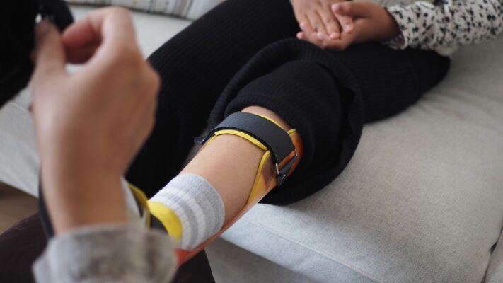
What Is an Orthosis? Why Is an Orthosis Used?
Ankara Ortopedi | Prof. Dr. Murat Demirel » General » What Is an Orthosis? Why [...]
13
Apr
Apr

Knee Replacement Surgery Prices
Ankara Ortopedi | Prof. Dr. Murat Demirel » Knee Treatments » Knee Replacement Surgery PricesThe [...]
18
Aug
Aug
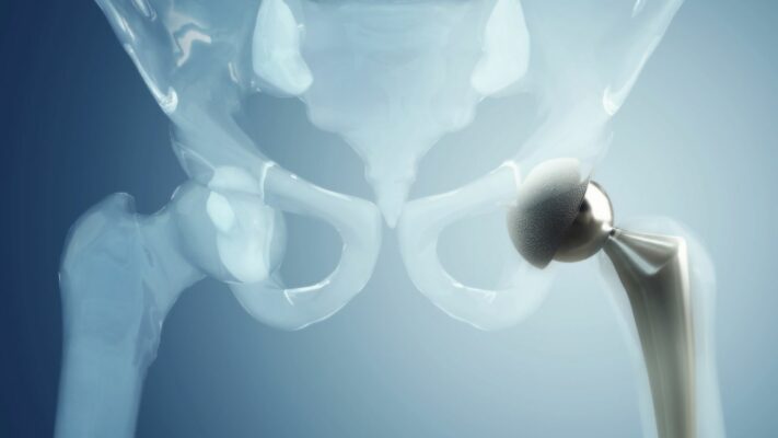
Hip Replacement Surgery Prices
Ankara Ortopedi | Prof. Dr. Murat Demirel » Hip Treatments » Hip Replacement Surgery PricesThe [...]
18
Aug
Aug